Explore
 Validate
 Learn
LearnPA5-54109
antibody from Invitrogen Antibodies
Targeting: PCNT
KEN, KIAA0402, PCN, PCNT2, PCNTB, SCKL4
 Immunocytochemistry
Antibody data
- Antibody Data
- Antigen structure
- References [1]
- Comments [0]
- Validations
- Immunocytochemistry [6]
- Other assay [1]
Submit
Validation data
Reference
Comment
Report error
- Product number
- PA5-54109 - Provider product page

- Provider
- Invitrogen Antibodies
- Product name
- Pericentrin Polyclonal Antibody
- Antibody type
- Polyclonal
- Antigen
- Recombinant protein fragment
- Description
- Immunogen sequence: MLDLSSWSSP EVLRKDWTLE PWPSLPVTPH SGALSLCSAD TSLGDRADTS LPQTQGPGLL CSPGVSAAAL ALQWAESPPA DDHHVQRTAV EKDVEDFITT SFDSQETLSS PPPGLEGKAD RSEKSDGSG Highest antigen sequence identity to the following orthologs: Mouse - 58%, Rat - 58%.
- Reactivity
- Human
- Host
- Rabbit
- Isotype
- IgG
- Vial size
- 100 μL
- Concentration
- 0.05 mg/mL
- Storage
- Store at 4°C short term. For long term storage, store at -20°C, avoiding freeze/thaw cycles.
Submitted references Meningioma cells express primary cilia but do not transduce ciliary Hedgehog signals.
Findakly S, Choudhury A, Daggubati V, Pekmezci M, Lang UE, Raleigh DR
Acta neuropathologica communications 2020 Jul 20;8(1):114
Acta neuropathologica communications 2020 Jul 20;8(1):114
No comments: Submit comment
Supportive validation
- Submitted by
- Invitrogen Antibodies (provider)
- Main image

- Experimental details
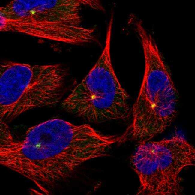
- Immunofluorescent staining of Pericentrin in human cell line U-251 MG shows positivity in centrosome. Samples were probed using a Pericentrin Polyclonal Antibody (Product # PA5-54109).
- Submitted by
- Invitrogen Antibodies (provider)
- Main image

- Experimental details
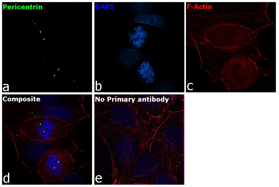
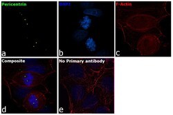
- Immunofluorescence analysis of Pericentrin was performed using MCF7 cells. The cells were fixed with 4% paraformaldehyde for 10 minutes, permeabilized with 0.1% Triton™ X-100 for 15 minutes, and blocked with 1% BSA for 1 hour at room temperature. The cells were labeled with Pericentrin Rabbit Polyclonal Antibody (Product # PA5-54109) at 4µg/mL in 0.1% BSA and incubated overnight at 4 degree and then labeled with Goat anti-Rabbit IgG (H+L) Superclonal™ Secondary Antibody, Alexa Fluor® 488 conjugate (Product # A27034) at a dilution of 1:2000 for 45 minutes at room temperature (Panel a: green). Nuclei (Panel b: blue) were stained with ProLong™ Diamond Antifade Mountant with DAPI (Product # P36962). F-actin (Panel c: red) was stained with Rhodamine Phalloidin (Product # R415, 1:300). Panel d represents the composite image showing centrosomal localization of Pericentrin. Panel e represents control cells with no primary antibody to assess background. The images were captured at 60X magnification. .
- Submitted by
- Invitrogen Antibodies (provider)
- Main image

- Experimental details
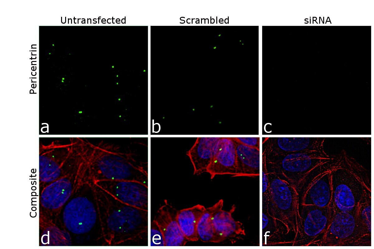
- KD of Pericentrin was achieved by transfecting MCF7 cells with Pericentrin specific siRNA (Silencer® select Product # s10136, s10137). Immunofluorescence analysis was performed on untransfected MCF7 cells (panel a,d), transfected with non-specific scrambled siRNA (panels b,e) and transfected with Pericentrin specific siRNA (panel c,f). Cells were fixed, permeabilized, and labelled with Pericentrin Rabbit Polyclonal Antibody (Product # PA5-54109, 4 µg/mL), followed by Goat anti-Rabbit IgG (H+L) Superclonal™ Secondary Antibody, Alexa Fluor® 488 conjugate (Product # A27034, 1:2000). Nuclei (blue) were stained using ProLong™ Diamond Antifade Mountant with DAPI (Product # P36962), and Rhodamine Phalloidin (Product # R415, 1:300) was used for cytoskeletal F-actin (red) staining. Reduction of specific signal was observed upon siRNA mediated KD (panel c,f) confirming specificity of the antibody to Pericentrin (green). The images were captured at 60X magnification..
- Submitted by
- Invitrogen Antibodies (provider)
- Main image

- Experimental details
- Immunofluorecent analysis of Pericentrin in human cell line U-251 MG using Pericentrin Polyclonal Antibody (Product # PA5-54109). Staining shows localization to centrosome.

- Submitted by
- Invitrogen Antibodies (provider)
- Main image

- Experimental details
- KD of Pericentrin was achieved by transfecting MCF7 cells with Pericentrin specific siRNA (Silencer® select Product # s10136, s10137). Immunofluorescence analysis was performed on untransfected MCF7 cells (panel a,d), transfected with non-specific scrambled siRNA (panels b,e) and transfected with Pericentrin specific siRNA (panel c,f). Cells were fixed, permeabilized, and labelled with Pericentrin Rabbit Polyclonal Antibody (Product # PA5-54109, 4 µg/mL), followed by Goat anti-Rabbit IgG (Heavy Chain) Superclonal™ Secondary Antibody, Alexa Fluor® 488 conjugate (Product # A27034, 1:2000). Nuclei (blue) were stained using ProLong™ Diamond Antifade Mountant with DAPI (Product # P36962), and Rhodamine Phalloidin (Product # R415, 1:300) was used for cytoskeletal F-actin (red) staining. Reduction of specific signal was observed upon siRNA mediated KD (panel c,f) confirming specificity of the antibody to Pericentrin (green). The images were captured at 60X magnification..

- Submitted by
- Invitrogen Antibodies (provider)
- Main image
- Experimental details
- Immunofluorescence analysis of Pericentrin was performed using MCF7 cells. The cells were fixed with 4% paraformaldehyde for 10 minutes, permeabilized with 0.1% Triton™ X-100 for 15 minutes, and blocked with 1% BSA for 1 hour at room temperature. The cells were labeled with Pericentrin Rabbit Polyclonal Antibody (Product # PA5-54109) at 4µg/mL in 0.1% BSA and incubated overnight at 4 degree and then labeled with Goat anti-Rabbit IgG (Heavy Chain) Superclonal™ Secondary Antibody, Alexa Fluor® 488 conjugate (Product # A27034) at a dilution of 1:2000 for 45 minutes at room temperature (Panel a: green). Nuclei (Panel b: blue) were stained with ProLong™ Diamond Antifade Mountant with DAPI (Product # P36962). F-actin (Panel c: red) was stained with Rhodamine Phalloidin (Product # R415, 1:300). Panel d represents the composite image showing centrosomal localization of Pericentrin. Panel e represents control cells with no primary antibody to assess background. The images were captured at 60X magnification. .

Supportive validation
- Submitted by
- Invitrogen Antibodies (provider)
- Main image
- Experimental details
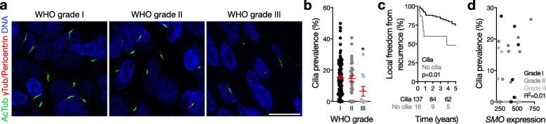
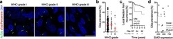
- Fig. 1 Meningiomas express primary cilia. a Confocal immunofluorescence microscopy for the ciliary marker acetylated tubulin (AcTub), the ciliary base/centriole markers gamma tubulin (gammaTub) and pericentrin, and DNA (DAPI) reveals that meningiomas express primary cilia. b Quantitative confocal immunofluorescence microscopy shows that WHO grade III meningiomas express less cilia than other meningiomas (* p = 0.04 ANOVA, p = 0.01 Student's t test). c Consistent with loss of cilia in high grade meningiomas, local freedom from recurrence is shorter in meningiomas without primary cilia compared to meningiomas expressing primary cilia (log-rank test). d Nanostring transcriptional profiling demonstrates that SMO expression does not correlate with cilia prevalence in meningioma