Explore
 Validate
 Learn
Learn Immunocytochemistry
Antibody data
- Antibody Data
- Antigen structure
- References [8]
- Comments [0]
- Validations
- Immunocytochemistry [1]
- Immunohistochemistry [1]
Submit
Validation data
Reference
Comment
Report error
- Product number
- HPA016820 - Provider product page

- Provider
- Atlas Antibodies
- Proper citation
- Atlas Antibodies Cat#HPA016820, RRID:AB_1855079
- Product name
- Anti-PCNT
- Antibody type
- Polyclonal
- Description
- Polyclonal Antibody against Human PCNT, Gene description: pericentrin, Alternative Gene Names: KEN, KIAA0402, PCN, PCNT2, PCNTB, SCKL4, Validated applications: ICC, IHC, Uniprot ID: O95613, Storage: Store at +4°C for short term storage. Long time storage is recommended at -20°C.
- Reactivity
- Human
- Host
- Rabbit
- Conjugate
- Unconjugated
- Isotype
- IgG
- Vial size
- 100 µl
- Concentration
- 0.3 mg/ml
- Storage
- Store at +4°C for short term storage. Long time storage is recommended at -20°C.
- Handling
- The antibody solution should be gently mixed before use.
Submitted references Joubert syndrome-derived induced pluripotent stem cells show altered neuronal differentiation in vitro
The Rab GTPase-binding protein EHBP1L1 and its interactors CD2AP/CIN85 negatively regulate the length of primary cilia via actin remodeling.
Essential role of hyperacetylated microtubules in innate immunity escape orchestrated by the EBV-encoded BHRF1 protein
A simple and fast method for fixation of cultured cell lines that preserves cellular structures containing gamma-tubulin.
Coupling bimolecular PARylation biosensors with genetic screens to identify PARylation targets.
Bisphenol A and its analogues disrupt centrosome cycle and microtubule dynamics in prostate cancer
Differing Effects of Herpes Simplex Virus 1 and Pseudorabies Virus Infections on Centrosomal Function
Antibody-based Protein Profiling of the Human Chromosome 21
De Mori R, Tardivo S, Pollara L, Giliani S, Ali E, Giordano L, Leuzzi V, Fischetto R, Gener B, Diprima S, Morelli M, Monti M, Sottile V, Valente E
Cell and Tissue Research 2024;396(2):255-267
Cell and Tissue Research 2024;396(2):255-267
The Rab GTPase-binding protein EHBP1L1 and its interactors CD2AP/CIN85 negatively regulate the length of primary cilia via actin remodeling.
Iwano T, Sobajima T, Takeda S, Harada A, Yoshimura SI
The Journal of biological chemistry 2023 Mar;299(3):102985
The Journal of biological chemistry 2023 Mar;299(3):102985
Essential role of hyperacetylated microtubules in innate immunity escape orchestrated by the EBV-encoded BHRF1 protein
Lin Z, Glon D, Vilmen G, Perdiz D, Hernandez E, Beauclair G, Quignon F, Berlioz-Torrent C, Maréchal V, Poüs C, Lussignol M, Esclatine A
PLOS Pathogens 2022;18(3):e1010371
PLOS Pathogens 2022;18(3):e1010371
A simple and fast method for fixation of cultured cell lines that preserves cellular structures containing gamma-tubulin.
Alvarado-Kristensson M
MethodsX 2018;5:227-233
MethodsX 2018;5:227-233
Coupling bimolecular PARylation biosensors with genetic screens to identify PARylation targets.
Krastev DB, Pettitt SJ, Campbell J, Song F, Tanos BE, Stoynov SS, Ashworth A, Lord CJ
Nature communications 2018 May 22;9(1):2016
Nature communications 2018 May 22;9(1):2016
Bisphenol A and its analogues disrupt centrosome cycle and microtubule dynamics in prostate cancer
Ho S, Rao R, To S, Schoch E, Tarapore P
Endocrine-Related Cancer 2017;24(2):83-96
Endocrine-Related Cancer 2017;24(2):83-96
Differing Effects of Herpes Simplex Virus 1 and Pseudorabies Virus Infections on Centrosomal Function
Pasdeloup D, Labetoulle M, Rixon F
Journal of Virology 2013;87(12):7102-7112
Journal of Virology 2013;87(12):7102-7112
Antibody-based Protein Profiling of the Human Chromosome 21
Uhlén M, Oksvold P, Älgenäs C, Hamsten C, Fagerberg L, Klevebring D, Lundberg E, Odeberg J, Pontén F, Kondo T, Sivertsson Å
Molecular & Cellular Proteomics 2012;11(3):M111.013458
Molecular & Cellular Proteomics 2012;11(3):M111.013458
No comments: Submit comment
Supportive validation
- Submitted by
- Atlas Antibodies (provider)
- Main image

- Experimental details
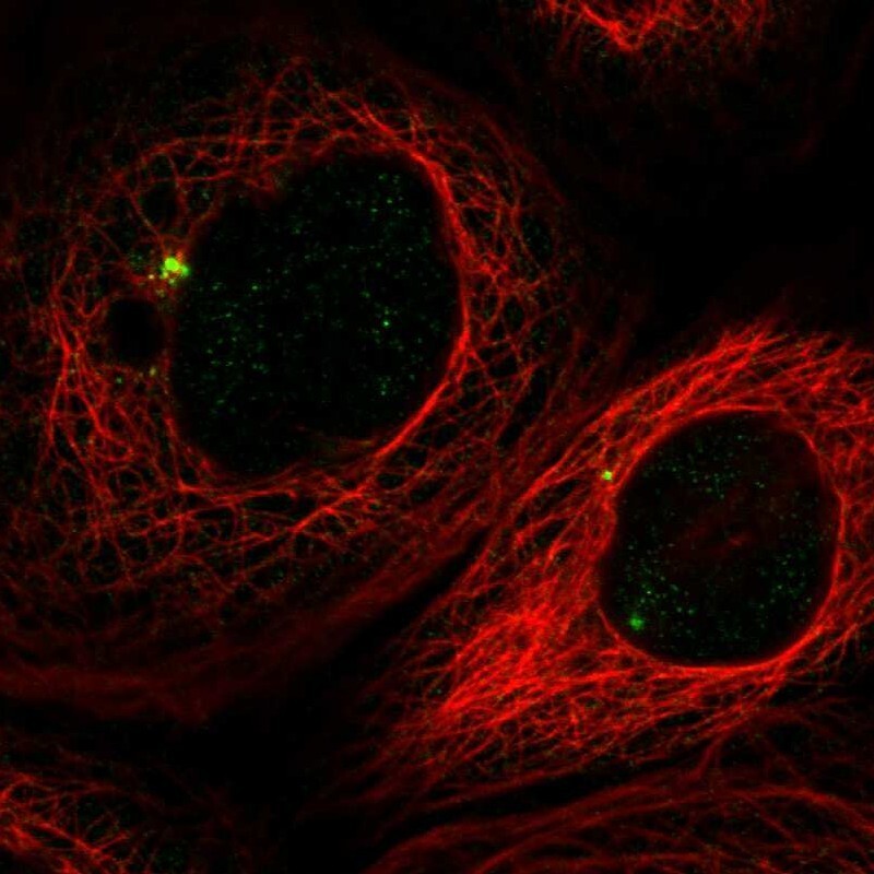
- Immunofluorescent staining of human cell line A-431 shows localization to centrosome.
- Sample type
- Human
Supportive validation
- Submitted by
- Atlas Antibodies (provider)
- Enhanced method
- Orthogonal validation
- Main image

- Experimental details
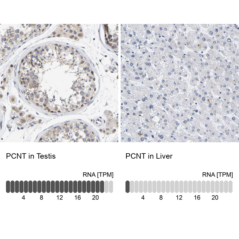
- Immunohistochemistry analysis in human testis and liver tissues using HPA016820 antibody. Corresponding PCNT RNA-seq data are presented for the same tissues.
- Sample type
- Human
- Protocol
- Protocol