Explore
 Validate
 Learn
Learn Western blot
 Immunohistochemistry
Antibody data
- Antibody Data
- Antigen structure
- References [1]
- Comments [0]
- Validations
- Immunohistochemistry [2]
- Other assay [3]
Submit
Validation data
Reference
Comment
Report error
- Product number
- PA5-54861 - Provider product page

- Provider
- Invitrogen Antibodies
- Product name
- RNF19A Polyclonal Antibody
- Antibody type
- Polyclonal
- Antigen
- Recombinant protein fragment
- Description
- Immunogen sequence: EMCTDKNSIF STNTSSDNGL TSISKQIGDF IECPLCLLRH SKDRFPDIMT CHHRSCVDCL Highest antigen sequence identity to the following orthologs: Mouse - 98%, Rat - 93%.
- Reactivity
- Human
- Host
- Rabbit
- Isotype
- IgG
- Vial size
- 100 μL
- Concentration
- 0.20 mg/mL
- Storage
- Store at 4°C short term. For long term storage, store at -20°C, avoiding freeze/thaw cycles.
Submitted references Ring finger protein 19A is overexpressed in non-small cell lung cancer and mediates p53 ubiquitin-degradation to promote cancer growth.
Cheng Y, Hu Y, Wang H, Zhao Z, Jiang X, Zhang Y, Zhang J, Tong Y, Qiu X
Journal of cellular and molecular medicine 2021 Aug;25(16):7796-7808
Journal of cellular and molecular medicine 2021 Aug;25(16):7796-7808
No comments: Submit comment
Supportive validation
- Submitted by
- Invitrogen Antibodies (provider)
- Main image
- Experimental details
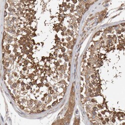
- Immunohistochemical staining of RNF19A in human testis using a RNF19A Polyclonal Antibody (Product # PA5-54861) shows strong cytoplasmic positivity in cells in seminiferous ducts.
- Submitted by
- Invitrogen Antibodies (provider)
- Main image

- Experimental details
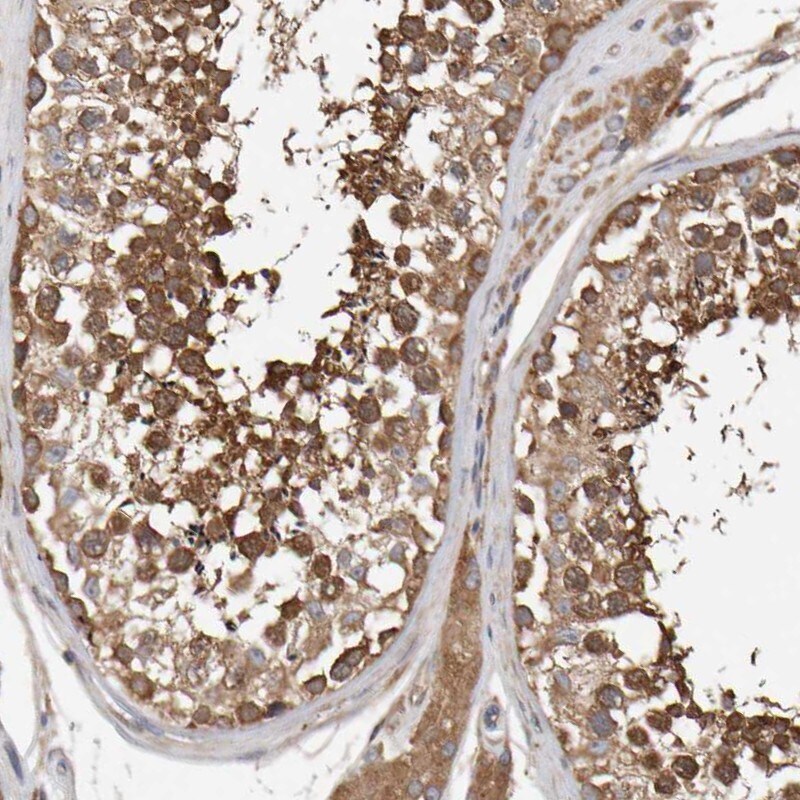
- Immunohistochemical analysis of RNF19A in human prostate using RNF19A Polyclonal Antibody (Product # PA5-54861) shows strong cytoplasmic positivity in smooth muscle cells.
Supportive validation
- Submitted by
- Invitrogen Antibodies (provider)
- Main image
- Experimental details
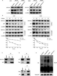
- FIGURE 5 RNF19A interacts with p53 and promotes p53 ubiquitination. A, RNF19A-silenced A549 and H460 cells were treated with MG132 and DMSO (control). RNF19A and p53 expression levels in the treated cells were detected via Western blotting. B, RNF19A-silenced A549 and H460 cells were treated with CHX at the indicated time points. RNF19A and p53 expression levels in the treated cells were detected via Western blotting and the half-life of p53 was calculated. C, Endogenous p53 and RNF19A form a protein complex in A549 cells. D, RNF19A co-immunoprecipitates with p53 in A549 cells transfected with p53 and FLAG-tagged RNF19A. E, A549 cells transfected with RNF19A expression plasmid or the RNF19A siRNA along with HA-ubiquitin (Ub). Levels of p53 ubiquitination were detected via immunoprecipitation using the anti-p53 antibody, followed by anti-HA immunoblotting

- Submitted by
- Invitrogen Antibodies (provider)
- Main image
- Experimental details
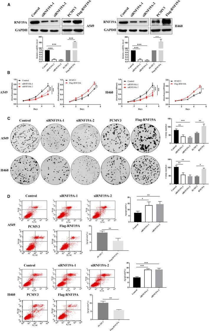
- FIGURE 2 RNF19A promotes NSCLC cell proliferation and inhibits apoptosis. A, RNF19A knockdown and overexpression was detected via Western blotting and qRT-PCR. B, Viability of transfected A549 and H460 cells was measured using the MTT assay. C, Colony formation was detected in transfected A549 and H460 cells. D, Transfected A549 and H460 cells were stained with Annexin V-FITC/PI. Stained cells were analysed via flow cytometry. * P < .05, ** P < .01 and *** P < .001. Data are presented as the mean +- SE of three independent experiments. SE, standard error
- Submitted by
- Invitrogen Antibodies (provider)
- Main image

- Experimental details
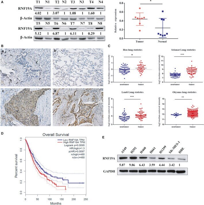
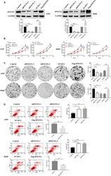
- FIGURE 1 RNF19A is highly expressed in NSCLC tissues and correlates with poor prognosis. A, Expression of RNF19A in eight matching NSCLC and non-tumour tissues was detected via Western blotting. T, tumour tissue; N, non-tumour tissue. * P < .05. B, IHC staining representative images of RNF19A in NSCLC and non-tumour specimens. a, bronchiole tissue; b, alveolar tissue; c, squamous cell carcinoma tissue; d, adenocarcinoma tissue. C, RNF19A mRNA expression comparison between NSCLC and non-tumour tissues from the Oncomine database. * P < .05, ** P < .01 and *** P < .001. D, RNF19A expression correlates with poor prognosis in patients with NSCLC, obtained from GEPIA data sets. * P < .05, ** P < .01 and *** P < .001. E, Immunoblot of RNF19A confirms high expression of RNF19A in NSCLC cell lines. IHC, immunohistochemistry; NSCLC, non-small cell lung cancer