 Explore
 Validate
 Learn
Learn Western blot
Antibody data
- Antibody Data
- Antigen structure
- References [0]
- Comments [0]
- Validations
- Western blot [1]
- Immunocytochemistry [2]
Submit
Validation data
Reference
Comment
Report error
- Product number
- LS-C774042 - Provider product page

- Provider
- LSBio
- Product name
- PPP5C Antibody (clone 12F7, Atto 633) LS-C774042
- Antibody type
- Monoclonal
- Description
- Protein G purified
- Reactivity
- Human
- Host
- Mouse
- Conjugate
- Red dye
- Isotype
- IgG
- Antibody clone number
- 12F7
- Storage
- Store at -20°C.
No comments: Submit comment
Supportive validation
- Submitted by
- LSBio (provider)
- Main image
- Experimental details
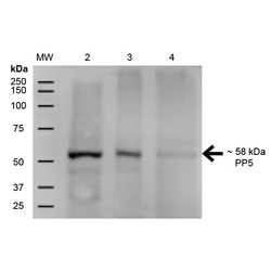
- Western Blot analysis of Human A431, HEK293, and Jurkat cell lysates showing detection of ~58 kDa PP5 protein using Mouse Anti-PP5 Monoclonal Antibody, Clone 12F7. Lane 1: MW Ladder. Lane 2: Human A431 (15 µg). Lane 3: Human HEK293 (15 µg). Lane 4: Human Jurkat (15 µg). Load: 15 µg. Block: 5% Skim Milk for 1 hour at RT. Primary Antibody: Mouse Anti-PP5 Monoclonal Antibody at 1:500 for 1 hour at RT. Secondary Antibody: Goat Anti-Mouse IgG: HRP at 1:200 for 1 hour at RT. Color Development: ECL solution for 6 min at RT. Predicted/Observed Size: ~58 kDa.
Supportive validation
- Submitted by
- LSBio (provider)
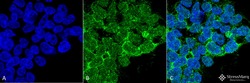
- Main image
- Experimental details
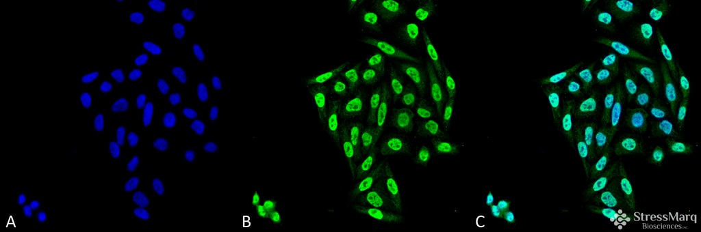
- Immunocytochemistry/Immunofluorescence analysis using Mouse Anti-PP5 Monoclonal Antibody, Clone 12F7. Tissue: Cervical cancer cell line (HeLa). Species: Human. Fixation: 4% Formaldehyde for 15 min at RT. Primary Antibody: Mouse Anti-PP5 Monoclonal Antibody at 1:100 for 60 min at RT. Secondary Antibody: Goat Anti-Mouse ATTO 488 at 1:100 for 60 min at RT. Counterstain: DAPI (blue) nuclear stain at 1:5000 for 5 min RT. Localization: Nucleus, Cytoplasm. Magnification: 40X.
- Submitted by
- LSBio (provider)
- Main image
- Experimental details
- Immunocytochemistry/Immunofluorescence analysis using Mouse Anti-PP5 Monoclonal Antibody, Clone 12F7. Tissue: Embryonic kidney epithelial cell line (HEK293). Species: Human. Fixation: 2% Formaldehyde for 20 min at RT. Primary Antibody: Mouse Anti-PP5 Monoclonal Antibody at 1:50 for 1 hour at RT. Secondary Antibody: Alexa Fluor 488 Goat Anti-Mouse (green) at 1:100 for 1 hour at RT. Counterstain: DAPI (blue) nuclear stain. Magnification: 63x.
