 Explore
 Validate
 Learn
Learn Western blot
 ELISA
ELISA Immunocytochemistry
Antibody data
- Antibody Data
- Antigen structure
- References [5]
- Comments [0]
- Validations
- Immunocytochemistry [3]
- Immunohistochemistry [1]
- Other assay [5]
Submit
Validation data
Reference
Comment
Report error
- Product number
- MA5-15502 - Provider product page

- Provider
- Invitrogen Antibodies
- Product name
- WNT5A Monoclonal Antibody (6F2)
- Antibody type
- Monoclonal
- Antigen
- Purifed from natural sources
- Description
- MA5-15502 targets WNT5A in indirect ELISA, IF, IHC, and WB applications and shows reactivity with Human samples. The MA5-15502 immunogen is purified recombinant fragment of WNT5A expressed in E. Coli.. MA5-15502 detects WNT5A which has a predicted molecular weight of approximately 42.3kDa.
- Reactivity
- Human
- Host
- Mouse
- Isotype
- IgG
- Antibody clone number
- 6F2
- Vial size
- 100 μL
- Concentration
- Conc. Not Determined
- Storage
- Store at 4°C short term. For long term storage, store at -20°C, avoiding freeze/thaw cycles.
Submitted references MiR-26a-5p as a useful therapeutic target for upper tract urothelial carcinoma by regulating WNT5A/β-catenin signaling.
Wnt5a promotes renal tubular inflammation in diabetic nephropathy by binding to CD146 through noncanonical Wnt signaling.
Ehrlichia chaffeensis TRP120 Is a Wnt Ligand Mimetic That Interacts with Wnt Receptors and Contains a Novel Repetitive Short Linear Motif That Activates Wnt Signaling.
Infant High-Grade Gliomas Comprise Multiple Subgroups Characterized by Novel Targetable Gene Fusions and Favorable Outcomes.
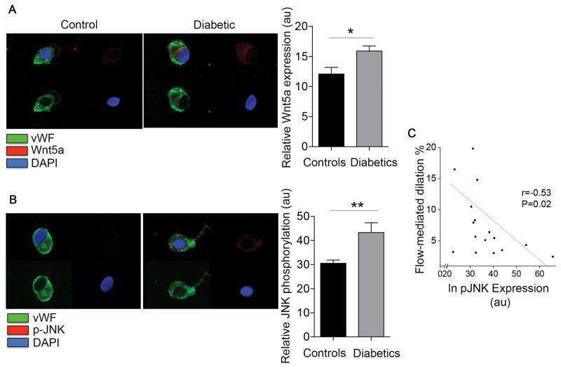
Endothelial Dysfunction in Human Diabetes Is Mediated by Wnt5a-JNK Signaling.
Chung YH, Cheng YT, Kao YH, Tsai WC, Huang GK, Chen YT, Shen YC, Tai MH, Chiang PH
Scientific reports 2022 Apr 28;12(1):6955
Scientific reports 2022 Apr 28;12(1):6955
Wnt5a promotes renal tubular inflammation in diabetic nephropathy by binding to CD146 through noncanonical Wnt signaling.
Li X, Wen J, Dong Y, Zhang Q, Guan J, Liu F, Zhou T, Li Z, Fan Y, Wang N
Cell death & disease 2021 Jan 18;12(1):92
Cell death & disease 2021 Jan 18;12(1):92
Ehrlichia chaffeensis TRP120 Is a Wnt Ligand Mimetic That Interacts with Wnt Receptors and Contains a Novel Repetitive Short Linear Motif That Activates Wnt Signaling.
Rogan MR, Patterson LL, Byerly CD, Luo T, Paessler S, Veljkovic V, Quade B, McBride JW
mSphere 2021 Apr 21;6(2)
mSphere 2021 Apr 21;6(2)
Infant High-Grade Gliomas Comprise Multiple Subgroups Characterized by Novel Targetable Gene Fusions and Favorable Outcomes.
Clarke M, Mackay A, Ismer B, Pickles JC, Tatevossian RG, Newman S, Bale TA, Stoler I, Izquierdo E, Temelso S, Carvalho DM, Molinari V, Burford A, Howell L, Virasami A, Fairchild AR, Avery A, Chalker J, Kristiansen M, Haupfear K, Dalton JD, Orisme W, Wen J, Hubank M, Kurian KM, Rowe C, Maybury M, Crosier S, Knipstein J, Schüller U, Kordes U, Kram DE, Snuderl M, Bridges L, Martin AJ, Doey LJ, Al-Sarraj S, Chandler C, Zebian B, Cairns C, Natrajan R, Boult JKR, Robinson SP, Sill M, Dunkel IJ, Gilheeney SW, Rosenblum MK, Hughes D, Proszek PZ, Macdonald TJ, Preusser M, Haberler C, Slavc I, Packer R, Ng HK, Caspi S, Popović M, Faganel Kotnik B, Wood MD, Baird L, Davare MA, Solomon DA, Olsen TK, Brandal P, Farrell M, Cryan JB, Capra M, Karremann M, Schittenhelm J, Schuhmann MU, Ebinger M, Dinjens WNM, Kerl K, Hettmer S, Pietsch T, Andreiuolo F, Driever PH, Korshunov A, Hiddingh L, Worst BC, Sturm D, Zuckermann M, Witt O, Bloom T, Mitchell C, Miele E, Colafati GS, Diomedi-Camassei F, Bailey S, Moore AS, Hassall TEG, Lowis SP, Tsoli M, Cowley MJ, Ziegler DS, Karajannis MA, Aquilina K, Hargrave DR, Carceller F, Marshall LV, von Deimling A, Kramm CM, Pfister SM, Sahm F, Baker SJ, Mastronuzzi A, Carai A, Vinci M, Capper D, Popov S, Ellison DW, Jacques TS, Jones DTW, Jones C
Cancer discovery 2020 Jul;10(7):942-963
Cancer discovery 2020 Jul;10(7):942-963
Endothelial Dysfunction in Human Diabetes Is Mediated by Wnt5a-JNK Signaling.
Bretón-Romero R, Feng B, Holbrook M, Farb MG, Fetterman JL, Linder EA, Berk BD, Masaki N, Weisbrod RM, Inagaki E, Gokce N, Fuster JJ, Walsh K, Hamburg NM
Arteriosclerosis, thrombosis, and vascular biology 2016 Mar;36(3):561-9
Arteriosclerosis, thrombosis, and vascular biology 2016 Mar;36(3):561-9
No comments: Submit comment
Supportive validation
- Submitted by
- Invitrogen Antibodies (provider)
- Main image
- Experimental details
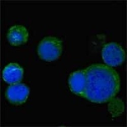
- Immunofluorescence analysis of PC-12 cells using WNT5A monoclonal antibody (Product # MA5-15502) (Green). Blue: DRAQ5 fluorescent DNA dye.

- Submitted by
- Invitrogen Antibodies (provider)
- Main image

- Experimental details
- Immunofluorescence analysis of PC-12 cells using WNT5A monoclonal antibody (Product # MA5-15502) (Green). Blue: DRAQ5 fluorescent DNA dye.

- Submitted by
- Invitrogen Antibodies (provider)
- Main image

- Experimental details
- Immunofluorescence analysis of PC-12 cells using WNT5A monoclonal antibody (Product # MA5-15502) (Green). Blue: DRAQ5 fluorescent DNA dye.

Supportive validation
- Submitted by
- Invitrogen Antibodies (provider)
- Main image

- Experimental details
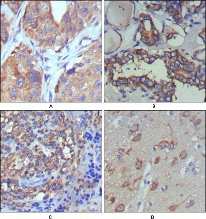
- Immunohistochemical analysis of paraffin-embedded human lung cancer (A), thyroid cancer (B), lymph node (C) and brain (D) showing cytoplasmic and extracellular matrix localization using WNT5A monoclonal antibody (Product # MA5-15502) followed with DAB staining.
Supportive validation
- Submitted by
- Invitrogen Antibodies (provider)
- Main image
- Experimental details
- NULL
- Submitted by
- Invitrogen Antibodies (provider)
- Main image
- Experimental details
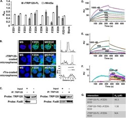
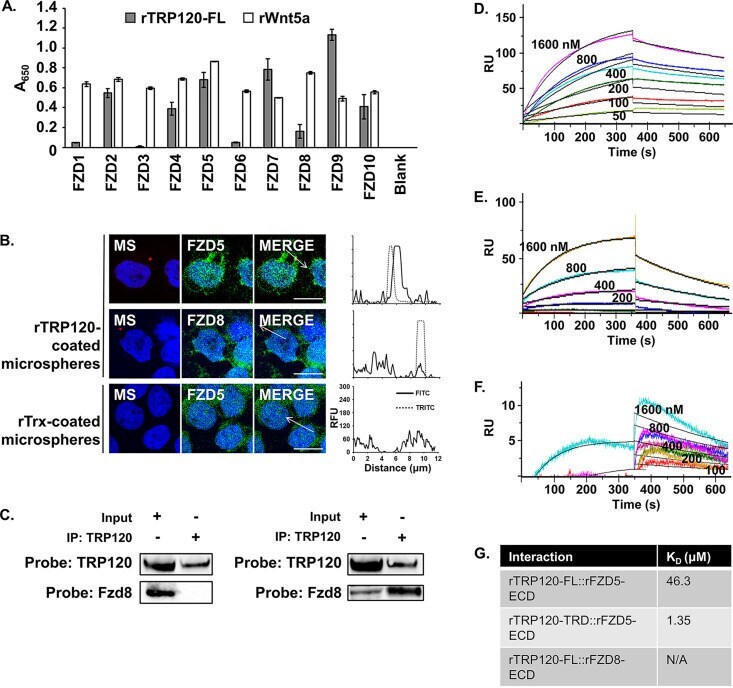
- FIG 2 TRP120 directly interacts with Fzd5. (A) rTRP120-FL interacts with rFzd2, 4, 5, 7, 9, and 10 ECD truncate proteins. To measure direct binding of TRP120 and Fzd proteins, recombinant Fzd extracellular domain (ECD) truncated proteins dissolved in PBS, or a blank, were adsorbed onto an ELISA plate and rTRP120-FL (gray) or rWnt5a (white) was added as an interacting protein. Positive interactions were detected by incubation with alpha-TRP120-I1 or alpha-Wnt5a followed by alkaline phosphatase-conjugated secondary antibodies. Data are an average from three independent experiments, and values represent the mean plus or minus standard deviation. (B) THP-1 cells were treated with rTrx- or rTRP120-FL-coated microspheres for 1 h. Cells were dual stained for coated microspheres (""MS,"" tetramethylrhodamine isothiocyanate [TRITC], red) or Fzd (fluorescein isothiocyanate [FITC], green), and nuclei were stained with DAPI (blue). Arrow represents dimensions of line profile graph of FITC channel (black line) and TRITC channel (dashed line). Scale bar = 10 mum. Images are representative of three independent experiments. (C) THP-1 cells were infected with E. chaffeensis (MOI 100), and whole-cell lysate was harvested 70 hpi. Lysate was run over immobilized alpha-TRP120, and the input lysate as well as the immunoprecipitated eluates was probed for Fzd5 and Fzd8 by immunoblotting. (D to F) One hundred nanomolar rTRP120-FL (D and F) or rTRP120-TRD (E) was immobilized on a Ni-nitrilotriacetic
- Submitted by
- Invitrogen Antibodies (provider)
- Main image
- Experimental details
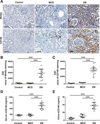
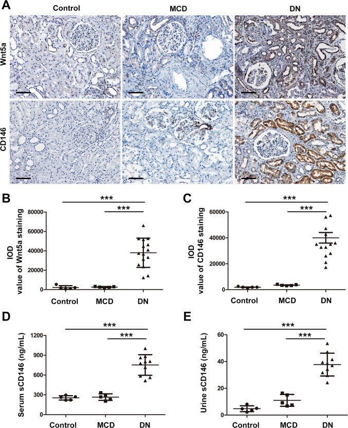
- Fig. 1 The expression levels of Wnt5a and CD146 were upregulated in diabetic neuropathy (DN) patients. A Representative immunostaining of Wnt5a and CD146 on normal kidney sections of nephrectomy samples ( n = 5) and kidney biopsy samples of patients with minimal change disease (MCD) ( n = 5) and DN ( n = 15). Original magnification, x200; scale bar, 100 mum. B Quantitative analysis of Wnt5a staining in kidney sections from each group according to the average integrated optical density (IOD). *** P < 0.001. C Quantitative analysis of expression of CD146 staining on kidney sections of each group by the average IOD. *** P < 0.001. D , E The concentration of soluble CD146 (sCD146) in serum and urine samples of each group. *** P < 0.001. Immunostaining was performed in duplicate. Significant differences were determined by unpaired two-tailed t -tests.
- Submitted by
- Invitrogen Antibodies (provider)
- Main image
- Experimental details
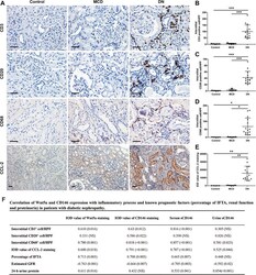
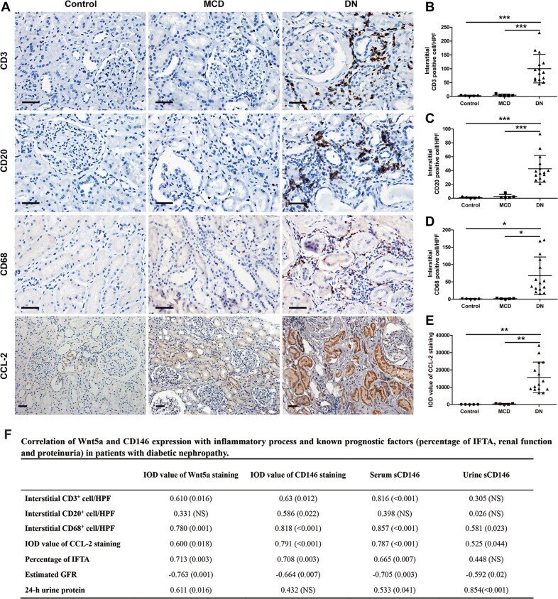
- Fig. 2 Assessment of renal inflammation in human kidney biopsies. A Immunostaining using specific antibodies for different immune cells, including T lymphocytes (CD3 + cells), B lymphocytes (CD20 + cells) and monocytes/macrophages (CD68 + cells), was performed on normal subjects, MCD patients, and DN patients. Original magnification, x400; scale bar, 50 mum. Immunohistochemical staining of the proinflammatory marker, chemokine (C-C motif) ligand 2 (CCL-2) was also performed. Original magnification, x200; scale bar, 50 mum. B - D The numbers of CD3 + , CD20 + , and CD68 + cells per high-power field (HPF) were counted in the tubulointerstitium. *** P < 0.001, * P < 0.05. E Quantitative analysis of the expression of CCL-2 staining on kidney sections of each group according to the average IOD. ** P < 0.01. Immunostaining was performed in duplicate. Significant differences were determined by unpaired two-tailed t -tests. F Correlation of Wnt5a and CD146 expression with inflammatory process and known prognostic factors (percentage of IFTA, renal function, and proteinuria) in patients with diabetic nephropathy. Data are r value based on Pearson and Spearman correlation analysis; ( P value); NS is not significant. IFTA, interstitial fibrosis and tubular atrophy; Estimated GFR, Estimated glomerular filtration rate; HbA1c, hemoglobin A1c.
- Submitted by
- Invitrogen Antibodies (provider)
- Main image
- Experimental details
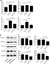
- Figure 6 miR-26a-5p overexpression modulated expression of Wnt5A/beta-catenin signaling mediators and downstream molecules in UTUC cells. BFTC-909 cells were transfected with miR-26a-5p mimics for 48 h and subjected to detection of mRNA and protein expression by using qPCR ( A ) and western blot ( B) , respectively. GAPDH was used as loading internal control. ( C ) The blotting densitometry of protein levels in BFTC-909 cells. Data are expressed as mean +- SEM (n = 3). *Indicates a P < 0.05 between the indicated groups.
