Explore
 Validate
 Learn
Learn Western blot
 Immunohistochemistry
Antibody data
- Antibody Data
- Antigen structure
- References [3]
- Comments [0]
- Validations
- Immunohistochemistry [1]
Submit
Validation data
Reference
Comment
Report error
- Product number
- HPA048533 - Provider product page

- Provider
- Atlas Antibodies
- Proper citation
- Atlas Antibodies Cat#HPA048533, RRID:AB_2680432
- Product name
- Anti-CYP17A1
- Antibody type
- Polyclonal
- Description
- Polyclonal Antibody against Human CYP17A1, Gene description: cytochrome P450, family 17, subfamily A, polypeptide 1, Alternative Gene Names: CPT7, CYP17, P450C17, S17AH, Validated applications: WB, IHC, Uniprot ID: P05093, Storage: Store at +4°C for short term storage. Long time storage is recommended at -20°C.
- Reactivity
- Human
- Host
- Rabbit
- Conjugate
- Unconjugated
- Isotype
- IgG
- Vial size
- 100 µl
- Concentration
- 0.1 mg/ml
- Storage
- Store at +4°C for short term storage. Long time storage is recommended at -20°C.
- Handling
- The antibody solution should be gently mixed before use.
Submitted references Steroid Profiles and Precursor-to-Product Ratios Are Altered in Pregnant Women with Preeclampsia
Accelerated telomere shortening in adrenal zona reticularis in patients with prolonged critical illness.
Biochemical, Histopathological, and Genetic Characterization of Posture-Responsive and Unresponsive APAs
Trummer O, Stern C, Reintar S, Mayer-Pickel K, Cervar-Zivkovic M, Dischinger U, Kurlbaum M, Huppertz B, Fluhr H, Obermayer-Pietsch B
International Journal of Molecular Sciences 2024;25(23):12704
International Journal of Molecular Sciences 2024;25(23):12704
Accelerated telomere shortening in adrenal zona reticularis in patients with prolonged critical illness.
Nonaka K, Takubo K, Aida J, Watai Y, Komatsu A, Gomi F, Shichi Y, Yamazaki Y, Ishiwata T, Sasano H, Arai T
Frontiers in endocrinology 2023;14:1244553
Frontiers in endocrinology 2023;14:1244553
Biochemical, Histopathological, and Genetic Characterization of Posture-Responsive and Unresponsive APAs
Guo Z, Nanba K, Udager A, McWhinney B, Ungerer J, Wolley M, Thuzar M, Gordon R, Rainey W, Stowasser M
The Journal of Clinical Endocrinology & Metabolism 2020;105(9):e3224-e3235
The Journal of Clinical Endocrinology & Metabolism 2020;105(9):e3224-e3235
No comments: Submit comment
Supportive validation
- Submitted by
- Atlas Antibodies (provider)
- Enhanced method
- Orthogonal validation
- Main image

- Experimental details
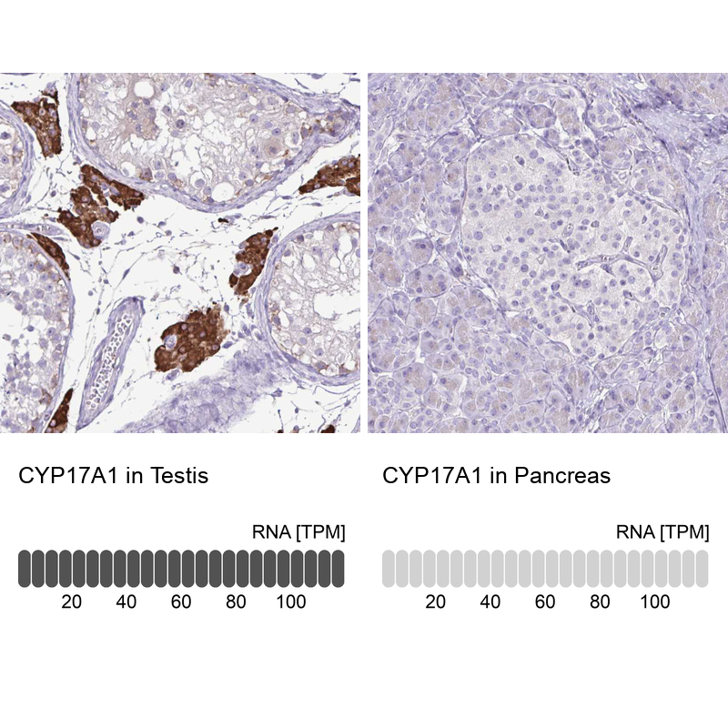
- Immunohistochemistry analysis in human testis and pancreas tissues using HPA048533 antibody. Corresponding CYP17A1 RNA-seq data are presented for the same tissues.
- Sample type
- Human
- Protocol
- Protocol