Explore
 Validate
 Learn
Learn Western blot
 Immunocytochemistry
 Immunoprecipitation
Antibody data
- Antibody Data
- Antigen structure
- References [3]
- Comments [0]
- Validations
- Immunocytochemistry [2]
- Other assay [2]
Submit
Validation data
Reference
Comment
Report error
- Product number
- 36-5100 - Provider product page

- Provider
- Invitrogen Antibodies
- Product name
- Connexin 31 Polyclonal Antibody
- Antibody type
- Polyclonal
- Antigen
- Synthetic peptide
- Description
- This antibody reacts with the ~32 kDa mouse Connexin 31 on Western blots; the band observed at ~62 kDa is probably due to dimerization of Connexin 31. Several higher bands of unknown origin are observed in western blots of mouse skin homogenates.
- Reactivity
- Human, Mouse
- Host
- Rabbit
- Isotype
- IgG
- Vial size
- 100 μg
- Concentration
- 0.25 mg/mL
- Storage
- Maintain refrigerated at 2-8°C for up to 1 month. For long term storage store at -20°C
Submitted references Comparative Analysis of Cx31 and Cx43 in Differentiation-Competent Rodent Keratinocytes.
Death of Neurons following Injury Requires Conductive Neuronal Gap Junction Channels but Not a Specific Connexin.
Connexin expression and gap junctional coupling in human cumulus cells: contribution to embryo quality.
Au A, Shao Q, White KK, Lucaciu SA, Esseltine JL, Barr K, Laird DW
Biomolecules 2020 Oct 14;10(10)
Biomolecules 2020 Oct 14;10(10)
Death of Neurons following Injury Requires Conductive Neuronal Gap Junction Channels but Not a Specific Connexin.
Fontes JD, Ramsey J, Polk JM, Koop A, Denisova JV, Belousov AB
PloS one 2015;10(5):e0125395
PloS one 2015;10(5):e0125395
Connexin expression and gap junctional coupling in human cumulus cells: contribution to embryo quality.
Wang HX, Tong D, El-Gehani F, Tekpetey FR, Kidder GM
Journal of cellular and molecular medicine 2009 May;13(5):972-84
Journal of cellular and molecular medicine 2009 May;13(5):972-84
No comments: Submit comment
Supportive validation
- Submitted by
- Invitrogen Antibodies (provider)
- Main image
- Experimental details
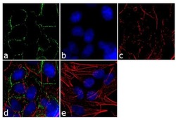
- Immunofluorescence analysis of CONNEXIN 31 was performed using 90% confluent log phase SH-SY5Y cells. The cells were fixed with 4% paraformaldehyde for 10 minutes, permeabilized with 0.1% Triton™ X-100 for 10 minutes, and blocked with 1% BSA for 1 hour at room temperature. The cells were labeled with Connexin 31 Rabbit Polyclonal Antibody (Product # 36-5100) at 2µg/mL in 0.1% BSA and incubated for 3 hours at room temperature and then labeled with Goat anti-Rabbit IgG (H+L) Superclonal™ Secondary Antibody, Alexa Fluor® 488 conjµgate (Product # A27034) at a dilution of 1:2000 for 45 minutes at room temperature (Panel a: green). Nuclei (Panel b: blue) were stained with SlowFade® Gold Antifade Mountant with DAPI (Product # S36938). F-actin (Panel c: red) was stained with Alexa Fluor® 555 Rhodamine Phalloidin (Product # R415, 1:300). Panel d represents the merged image showing cell junction localization. Panel e shows the no primary antibody control. The images were captured at 60X magnification.

- Submitted by
- Invitrogen Antibodies (provider)
- Main image

- Experimental details
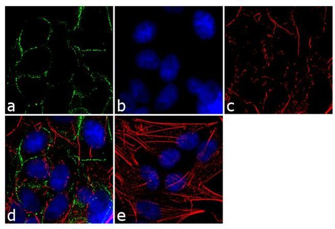
- Immunofluorescence analysis of CONNEXIN 31 was performed using 90% confluent log phase SH-SY5Y cells. The cells were fixed with 4% paraformaldehyde for 10 minutes, permeabilized with 0.1% Triton™ X-100 for 10 minutes, and blocked with 1% BSA for 1 hour at room temperature. The cells were labeled with Connexin 31 Rabbit Polyclonal Antibody (Product # 36-5100) at 2µg/mL in 0.1% BSA and incubated for 3 hours at room temperature and then labeled with Goat anti-Rabbit IgG (Heavy Chain) Superclonal™ Secondary Antibody, Alexa Fluor® 488 conjµgate (Product # A27034) at a dilution of 1:2000 for 45 minutes at room temperature (Panel a: green). Nuclei (Panel b: blue) were stained with SlowFade® Gold Antifade Mountant with DAPI (Product # S36938). F-actin (Panel c: red) was stained with Alexa Fluor® 555 Rhodamine Phalloidin (Product # R415, 1:300). Panel d represents the merged image showing cell junction localization. Panel e shows the no primary antibody control. The images were captured at 60X magnification.
Supportive validation
- Submitted by
- Invitrogen Antibodies (provider)
- Main image
- Experimental details
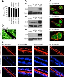
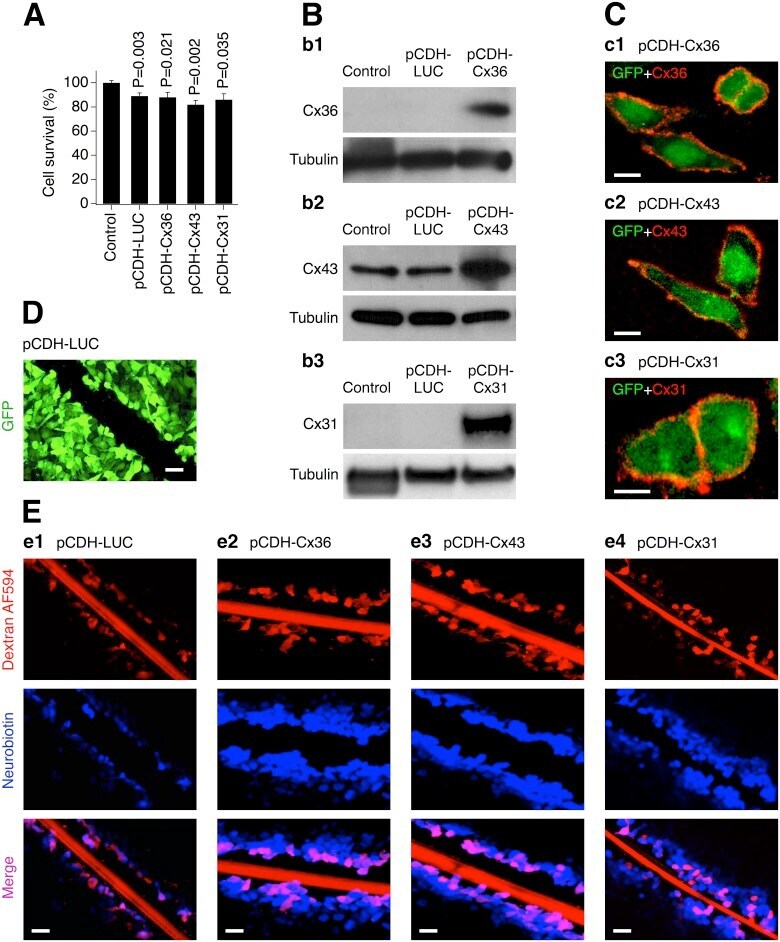
- Fig 2 Expression of connexins from lentivirus induces functional gap junction channels. Data from MTT assay ( A ), western blot ( B ), immunostaining ( C ) and scrape-loading dye transfer ( D, E ) experiments in transduced Cx36 knockout mouse neuronal cortical cultures ( A , B ) and stably-transduced HeLa cells ( C-E ) are shown. A , Lentiviral transductions reduce neuronal survival over a three day period (statistical analysis: Student's t -test, shown relative to non-infected {Control} cultures; n = 5-10 per group; mean +- SEM). However, no statistical difference in neuronal survival between the control lentivirus and each of the other transductions was detected (P>0.05, not shown; Student's t -test). B , Lentiviral transductions express Cx36 ( b1 ), Cx43 ( b2 ) and Cx31 ( b3 ) (these neuronal cultures have contained small amount of astrocytes that explains some background expression of Cx43). In A , B , cultures were infected on DIV6 and the analyses were done three ( A ) and two ( B ) days post-transduction. C , Connexins are expressed in the cell's plasma membrane ( c1-c3 ). Shown are superimposed confocal images of GFP (green; denotes the transduced cells) and staining for the corresponding connexin (red). D , This image illustrates that HeLa cells stably-transduced with pCDH-LUC all express GFP (the same was observed for all three connexin-expressing lentiviruses, but is not shown here). The image was taken from the same microscope field as in e1 (see be
- Submitted by
- Invitrogen Antibodies (provider)
- Main image
- Experimental details
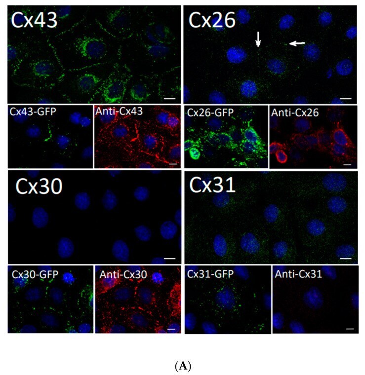
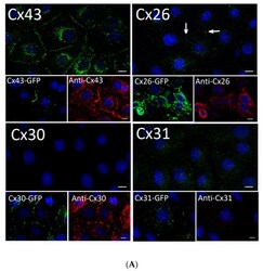
- Figure 2 Rat epidermal keratinocytes (REKs) express endogenous Cx43 and retain the capacity to assemble GFP-tagged Cx26, Cx30 and Cx31 into gap junctions. ( A ) Basal keratinocyte-like REKs express endogenous Cx43, trace amounts of Cx26 (arrows) but no detectable levels of Cx30 or Cx31 protein. REKs engineered to express GFP-tagged connexins were used to demonstrate that the anti-connexin antibodies were efficient at detecting connexins with the exception of the anti-Cx31 antibody. ( B ) REKs engineered to express GFP-tagged Cx43, Cx26, Cx30 and Cx31 all assembled prototypical gap junctions characterized by small punctate structures at sites of cell-cell interface (arrows). In addition, an array of intracellular fluorescent structures could be identified that reflect organelles and vesicles involved in connexin transport. Nuclei were stained with Hoechst dye. Bars = 10 mum.