Explore
 Validate
 Learn
Learn Western blot
Antibody data
- Antibody Data
- Antigen structure
- References [0]
- Comments [0]
- Validations
- Western blot [2]
- Immunocytochemistry [1]
- Immunohistochemistry [14]
Submit
Validation data
Reference
Comment
Report error
- Product number
- MA5-27191 - Provider product page

- Provider
- Invitrogen Antibodies
- Product name
- MEIS1 Monoclonal Antibody (OTI2A3)
- Antibody type
- Monoclonal
- Antigen
- Recombinant protein fragment
- Reactivity
- Human, Mouse, Rat
- Host
- Mouse
- Isotype
- IgG
- Antibody clone number
- OTI2A3
- Vial size
- 100 µL
- Concentration
- 1 mg/mL
- Storage
- -20° C, Avoid Freeze/Thaw Cycles
No comments: Submit comment
Supportive validation
- Submitted by
- Invitrogen Antibodies (provider)
- Main image

- Experimental details
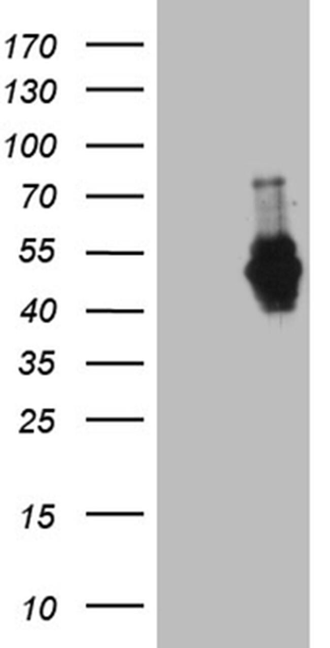
- Western blot analysis of MEIS1 in HEK293T cells in untransfected (Left lane) and transfected (Right lane) samples using 5 µg per lane. The samples were separated by SDS-PAGE and probed with MEIS1 (Product # MA5-27191) monoclonal antibody.
- Submitted by
- Invitrogen Antibodies (provider)
- Main image
- Experimental details
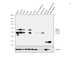
- Western blot was performed using Anti-MEIS1 Monoclonal Antibody (Product # MA5-27191). A 50 kDa band and a 41kDa band corresponding to different isoforms of MEIS1 was observed in IMR-32, SK-N-AS, K-562, HeLa cell lines. The 50 kDa band corresponding to MEIS1 was also observed in Mouse Cerebellum, Mouse Lung, Rat Lung and Mouse Skeletal muscle. A non-characterized band corresponding to 58 kDa was observed in the cell lines tested. Modified Whole cell extracts (30 µg lysate) of IMR-32 (Lane 1), SK-A-NS (Lane 2), K-562 (Lane 3), Jurkat (Lane 4), HeLa (Lane 5), Daudi (Lane 6), tissue extracts of Mouse Cerebellum (Lane 7), Mouse Lung (Lane 8), Rat Lung (Lane 9), Mouse Skeletal Muscle (lane 10) and Mouse Spleen (Lane 11) were electrophoresed using NuPAGE™ 4-12% Bis-Tris Protein Gel (Product # NP0322BOX). Resolved proteins were then transferred onto a nitrocellulose membrane (Product # IB23001) by iBlot® 2 Dry Blotting System (Product # IB21001). The blot was probed with the primary antibody (1:500 dilution) and detected by chemiluminescence Goat Anti-Mouse IgG Secondary Antibody, HRP conjugate (Product # A28177, 1:4000 dilution) using the iBright FL 1000 (Product # A32752). Chemiluminescent detection was performed using Novex® ECL Chemiluminescent Substrate Reagent Kit (Product # WP20005).

Supportive validation
- Submitted by
- Invitrogen Antibodies (provider)
- Main image
- Experimental details
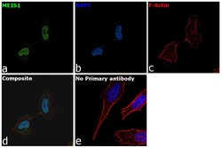
- Immunofluorescence analysis of MEIS1 was performed using 70% confluent log phase HeLa cells. The cells were fixed with 4% Paraformaldehyde for 10 minutes, permeabilized with 0.1% Triton™ X-100 for 15 minutes, and blocked with 2% BSA for 10 minutes at room temperature. The cells were labeled with MEIS1 Monoclonal Antibody (Product # MA5-27191) at 1:500 dilution in 0.1% BSA, incubated at 4 degree Celsius overnight and then labeled with Goat anti-Mouse IgG (H+L) Superclonal™ Secondary Antibody, Alexa Fluor® 488 conjugate (Product # A28175) at a dilution of 1:2000 dilution for 45 minutes at room temperature (Panel a: Green). Nuclei (Panel b: Blue) were stained with ProLong™ Diamond Antifade Mountant with DAPI (Product # P36962). F-actin (Panel c: Red) was stained with Rhodamine Phalloidin (Product # R415, 1:300). Panel d represents the merged image showing Cytoplasmic localization. Panel e represents control cells with no primary antibody to assess background. The images were captured at 60X magnification.

Supportive validation
- Submitted by
- Invitrogen Antibodies (provider)
- Main image

- Experimental details
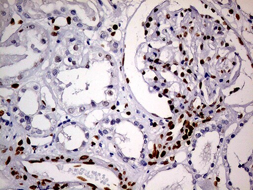
- Immunohistochemistry was performed on paraffin-embedded human kidney tissue. To expose target proteins, heat-induced epitope retrieval by 10mM citric buffer, pH6.0, 100°C for 10min. Following antigen retrieval, tissues were probed with a MEIS1 monoclonal antibody (Product # MA5-27191) at a dilution of 1:500.
- Submitted by
- Invitrogen Antibodies (provider)
- Main image

- Experimental details
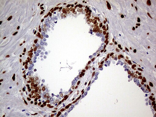
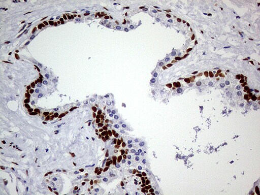
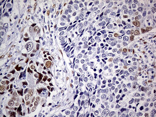
- Immunohistochemistry was performed on paraffin-embedded carcinoma of human prostate tissue. To expose target proteins, heat-induced epitope retrieval by 10mM citric buffer, pH6.0, 100°C for 10min. Following antigen retrieval, tissues were probed with a MEIS1 monoclonal antibody (Product # MA5-27191) at a dilution of 1:500.
- Submitted by
- Invitrogen Antibodies (provider)
- Main image

- Experimental details
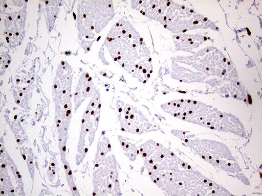
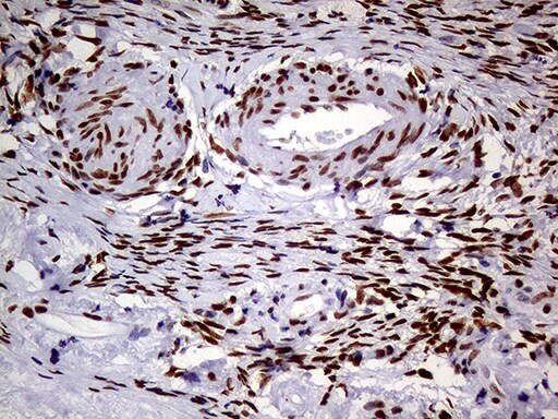
- Immunohistochemistry was performed on paraffin-embedded human bladder tissue. To expose target proteins, heat-induced epitope retrieval by 10mM citric buffer, pH6.0, 100°C for 10min. Following antigen retrieval, tissues were probed with a MEIS1 monoclonal antibody (Product # MA5-27191) at a dilution of 1:500.
- Submitted by
- Invitrogen Antibodies (provider)
- Main image
- Experimental details
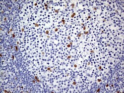
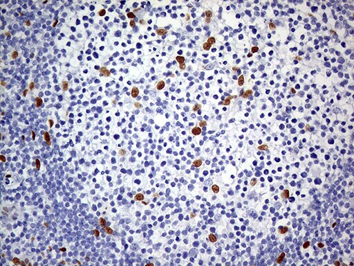
- Immunohistochemistry was performed on paraffin-embedded human lymph node tissue. To expose target proteins, heat-induced epitope retrieval by 10mM citric buffer, pH6.0, 100°C for 10min. Following antigen retrieval, tissues were probed with a MEIS1 monoclonal antibody (Product # MA5-27191) at a dilution of 1:500.
- Submitted by
- Invitrogen Antibodies (provider)
- Main image

- Experimental details
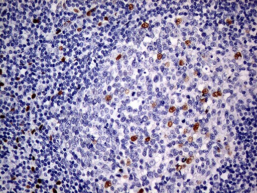
- Immunohistochemistry was performed on paraffin-embedded human tonsil tissue. To expose target proteins, heat-induced epitope retrieval by 10mM citric buffer, pH6.0, 100°C for 10min. Following antigen retrieval, tissues were probed with a MEIS1 monoclonal antibody (Product # MA5-27191) at a dilution of 1:500.
- Submitted by
- Invitrogen Antibodies (provider)
- Main image

- Experimental details
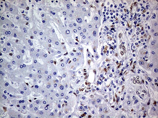
- Immunohistochemistry was performed on paraffin-embedded carcinoma of human liver tissue. To expose target proteins, heat-induced epitope retrieval by 10mM citric buffer, pH6.0, 100°C for 10min. Following antigen retrieval, tissues were probed with a MEIS1 monoclonal antibody (Product # MA5-27191) at a dilution of 1:500.
- Submitted by
- Invitrogen Antibodies (provider)
- Main image
- Experimental details
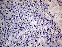
- Immunohistochemistry was performed on paraffin-embedded human liver tissue. To expose target proteins, heat-induced epitope retrieval by 1mM EDTA in 10mM Tris buffer (pH8.5) at 120°C for 3 min. Following antigen retrieval, tissues were probed with a MEIS1 monoclonal antibody (Product # MA5-27191) at a dilution of 1:500.

- Submitted by
- Invitrogen Antibodies (provider)
- Main image

- Experimental details
- Immunohistochemistry was performed on paraffin-embedded carcinoma of human lung tissue. To expose target proteins, heat-induced epitope retrieval by 10mM citric buffer, pH6.0, 100°C for 10min. Following antigen retrieval, tissues were probed with a MEIS1 monoclonal antibody (Product # MA5-27191) at a dilution of 1:500.
- Submitted by
- Invitrogen Antibodies (provider)
- Main image

- Experimental details
- Immunohistochemistry was performed on paraffin-embedded human ovary tissue. To expose target proteins, heat-induced epitope retrieval by 10mM citric buffer, pH6.0, 100°C for 10min. Following antigen retrieval, tissues were probed with a MEIS1 monoclonal antibody (Product # MA5-27191) at a dilution of 1:500.
- Submitted by
- Invitrogen Antibodies (provider)
- Main image

- Experimental details
- Immunohistochemistry was performed on paraffin-embedded adenocarcinoma of human ovary tissue. To expose target proteins, heat-induced epitope retrieval by 10mM citric buffer, pH6.0, 100°C for 10min. Following antigen retrieval, tissues were probed with a MEIS1 monoclonal antibody (Product # MA5-27191) at a dilution of 1:500.
- Submitted by
- Invitrogen Antibodies (provider)
- Main image

- Experimental details
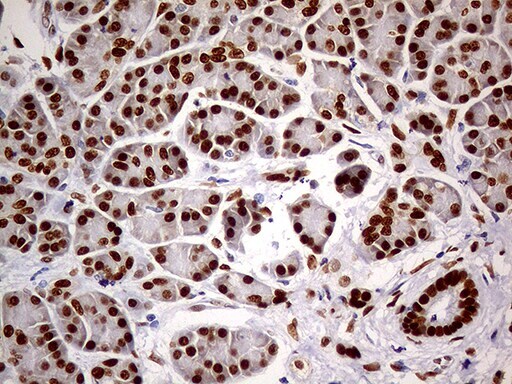
- Immunohistochemistry was performed on paraffin-embedded human pancreas tissue. To expose target proteins, heat-induced epitope retrieval by 10mM citric buffer, pH6.0, 100°C for 10min. Following antigen retrieval, tissues were probed with a MEIS1 monoclonal antibody (Product # MA5-27191) at a dilution of 1:500.
- Submitted by
- Invitrogen Antibodies (provider)
- Main image

- Experimental details
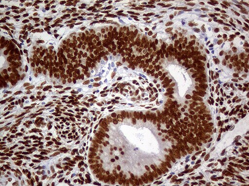
- Immunohistochemistry was performed on paraffin-embedded human endometrium tissue. To expose target proteins, heat-induced epitope retrieval by 10mM citric buffer, pH6.0, 100°C for 10min. Following antigen retrieval, tissues were probed with a MEIS1 monoclonal antibody (Product # MA5-27191) at a dilution of 1:500.
- Submitted by
- Invitrogen Antibodies (provider)
- Main image

- Experimental details
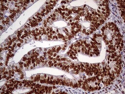
- Immunohistochemistry was performed on paraffin-embedded adenocarcinoma of human endometrium tissue. To expose target proteins, heat-induced epitope retrieval by 10mM citric buffer, pH6.0, 100°C for 10min. Following antigen retrieval, tissues were probed with a MEIS1 monoclonal antibody (Product # MA5-27191) at a dilution of 1:500.
- Submitted by
- Invitrogen Antibodies (provider)
- Main image

- Experimental details
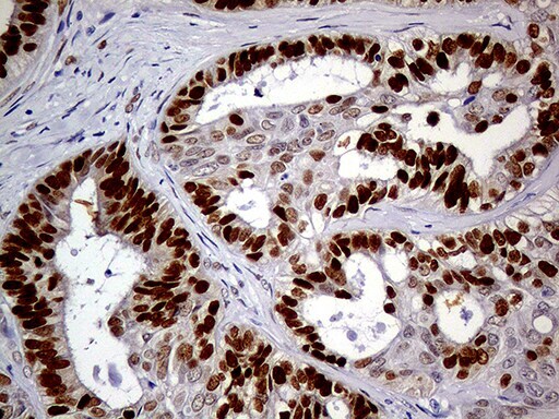
- Immunohistochemistry was performed on paraffin-embedded human prostate tissue. To expose target proteins, heat-induced epitope retrieval by 10mM citric buffer, pH6.0, 100°C for 10min. Following antigen retrieval, tissues were probed with a MEIS1 monoclonal antibody (Product # MA5-27191) at a dilution of 1:500.