 Explore
 Validate
 Learn
Learn Western blot
 ELISA
ELISA Immunoprecipitation
 Immunohistochemistry
Antibody data
- Antibody Data
- Antigen structure
- References [0]
- Comments [0]
- Validations
- Western blot [4]
- Immunoprecipitation [4]
Submit
Validation data
Reference
Comment
Report error
- Product number
- LS-C678638 - Provider product page

- Provider
- LSBio
- Product name
- MEIS1 Antibody LS-C678638
- Antibody type
- Polyclonal
- Description
- Protein G purified
- Reactivity
- Human
- Host
- Rabbit
- Isotype
- IgG
- Storage
- Upon receipt, store at -20°C or -80°C. Avoid repeated freeze.
No comments: Submit comment
Enhanced validation
- Submitted by
- LSBio (provider)
- Enhanced method
- Genetic validation
- Main image
- Experimental details
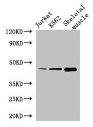
- Western Blot Positive WB detected in: Jurkat whole cell lysate, K562 whole cell lysate, Mouse skeletal muscle tissue All lanes: MEIS1 antibody at 2.8µg/ml Secondary Goat polyclonal to rabbit IgG at 1/50000 dilution Predicted band size: 44, 51 kDa Observed band size: 44 kDa

- Submitted by
- LSBio (provider)
- Enhanced method
- Genetic validation
- Main image

- Experimental details
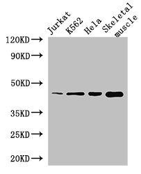
- Western Blot Positive WB detected in:Jurkat whole cell lysate,K562 whole cell lysate,Hela whole cell lysate,Mouse skeletal muscle tissue All Lanes: MEIS1 antibody at 2.8ug/ml Secondary Goat polyclonal to rabbit IgG at 1/50000 dilution Predicted band size: 44,51 kDa Observed band size: 44 kDa
- Submitted by
- LSBio (provider)
- Main image

- Experimental details
- Western Blot Positive WB detected in:Jurkat whole cell lysate,K562 whole cell lysate,Hela whole cell lysate,Mouse skeletal muscle tissue All Lanes: MEIS1 antibody at 2.8ug/ml Secondary Goat polyclonal to rabbit IgG at 1/50000 dilution Predicted band size: 44,51 kDa Observed band size: 44 kDa

- Submitted by
- LSBio (provider)
- Main image

- Experimental details
- Western Blot Positive WB detected in: Jurkat whole cell lysate, K562 whole cell lysate, Mouse skeletal muscle tissue All lanes: MEIS1 antibody at 2.8µg/ml Secondary Goat polyclonal to rabbit IgG at 1/50000 dilution Predicted band size: 44, 51 kDa Observed band size: 44 kDa

Supportive validation
- Submitted by
- LSBio (provider)
- Enhanced method
- Genetic validation
- Main image

- Experimental details
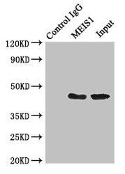
- Immunoprecipitating MEIS1 in K562 whole cell lysate Lane 1: Rabbit monoclonal IgG (1µg) instead of product in K562 whole cell lysate.For western blotting,a HRP-conjugated Protein G antibody was used as the Secondary antibody (1/50000) Lane 2: product (4µg) + K562 whole cell lysate (500µg) Lane 3: K562 whole cell lysate (20µg)

- Submitted by
- LSBio (provider)
- Main image

- Experimental details
- Immunoprecipitating MEIS1 in K562 whole cell lysate Lane 1: Rabbit monoclonal IgG (1µg) instead of product in K562 whole cell lysate.For western blotting,a HRP-conjugated Protein G antibody was used as the Secondary antibody (1/50000) Lane 2: product (4µg) + K562 whole cell lysate (500µg) Lane 3: K562 whole cell lysate (20µg)

- Submitted by
- LSBio (provider)
- Main image
- Experimental details
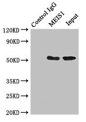
- Immunoprecipitating MEIS1 in K562 whole cell lysate Lane 1: Rabbit monoclonal IgG (1µg) instead of MEIS1 Antibody in K562 whole cell lysate.For western blotting, a HRP-conjugated Protein G antibody was used as the secondary antibody (1/50000) Lane 2: MEIS1 Antibody (4µg) + K562 whole cell lysate (500µg) Lane 3: K562 whole cell lysate (20µg)

- Submitted by
- LSBio (provider)
- Main image
- Experimental details
- Immunoprecipitating MEIS1 in K562 whole cell lysate Lane 1: Rabbit monoclonal IgG (1µg) instead of product in K562 whole cell lysate.For western blotting,a HRP-conjugated Protein G antibody was used as the Secondary antibody (1/50000) Lane 2: product (4µg) + K562 whole cell lysate (500µg) Lane 3: K562 whole cell lysate (20µg)
