Explore
 Validate
 Learn
Learn Western blot
 Immunohistochemistry
Antibody data
- Antibody Data
- Antigen structure
- References [0]
- Comments [0]
- Validations
- Western blot [1]
- Immunocytochemistry [3]
Submit
Validation data
Reference
Comment
Report error
- Product number
- PA5-77167 - Provider product page

- Provider
- Invitrogen Antibodies
- Product name
- PHIP Polyclonal Antibody
- Antibody type
- Polyclonal
- Antigen
- Recombinant full-length protein
- Description
- The antibody was affinity-purified from rabbit antiserum by affinity-chromatography using epitope-specific immunogen and the purity is > 95% (by SDS-PAGE).
- Reactivity
- Human, Mouse
- Host
- Rabbit
- Isotype
- IgG
- Vial size
- 100 µL
- Concentration
- 1 mg/mL
- Storage
- Store at 4°C short term. For long term storage, store at -20°C, avoiding freeze/thaw cycles.
No comments: Submit comment
Supportive validation
- Submitted by
- Invitrogen Antibodies (provider)
- Main image
- Experimental details
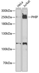
- Western blot analysis of PHIP in extracts of various cell lines. Samples were incubated with PHIP polyclonal antibody (Product # PA5-77167).

Supportive validation
- Submitted by
- Invitrogen Antibodies (provider)
- Main image
- Experimental details
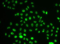
- Immunofluorescence analysis of PHIP in MCF7 cells. Samples were incubated with PHIP polyclonal antibody (Product # PA5-77167).
- Submitted by
- Invitrogen Antibodies (provider)
- Main image

- Experimental details
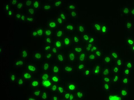
- Immunofluorescence analysis of PH-interacting protein (PHIP) was performed using 70% confluent log phase A-431 and U-87 MG cells. The cells were fixed with 4% paraformaldehyde for 10 minutes, permeabilized with 0.1% Triton™ X-100 for 10 minutes, and blocked with 2% BSA for 1 hour at room temperature. The cells were labeled with PHIP Polyclonal Antibody (Product # PA5-77167) at 1:100 in 0.1% BSA, incubated at 4 degree celsius overnight and then labeled with Goat anti-Rabbit IgG (H+L) Highly Cross-Adsorbed Secondary Antibody, Alexa Fluor Plus 488 (Product # A32731), (1:2500), for 45 minutes at room temperature (Panel a: Green). Nuclei (Panel b:Blue) were stained with Hoechst 33342 (Product # H1399). F-actin (Panel c: Red) was stained with Rhodamine Phalloidin (Product # R415, 1:300). Panel d represents the merged image showing nuclear localization for PHIP in A-431 and low to no signal in U-87 MG (Panel e) which is reported low expressing for the protein. Panel f represents control A-431 cells with no primary antibody to assess background. The images were captured at 40X magnification with CellInsight CX7 LZR High-Content Screening (HCS) Platform (Product # CX7C1115LZR).

- Submitted by
- Invitrogen Antibodies (provider)
- Main image

- Experimental details
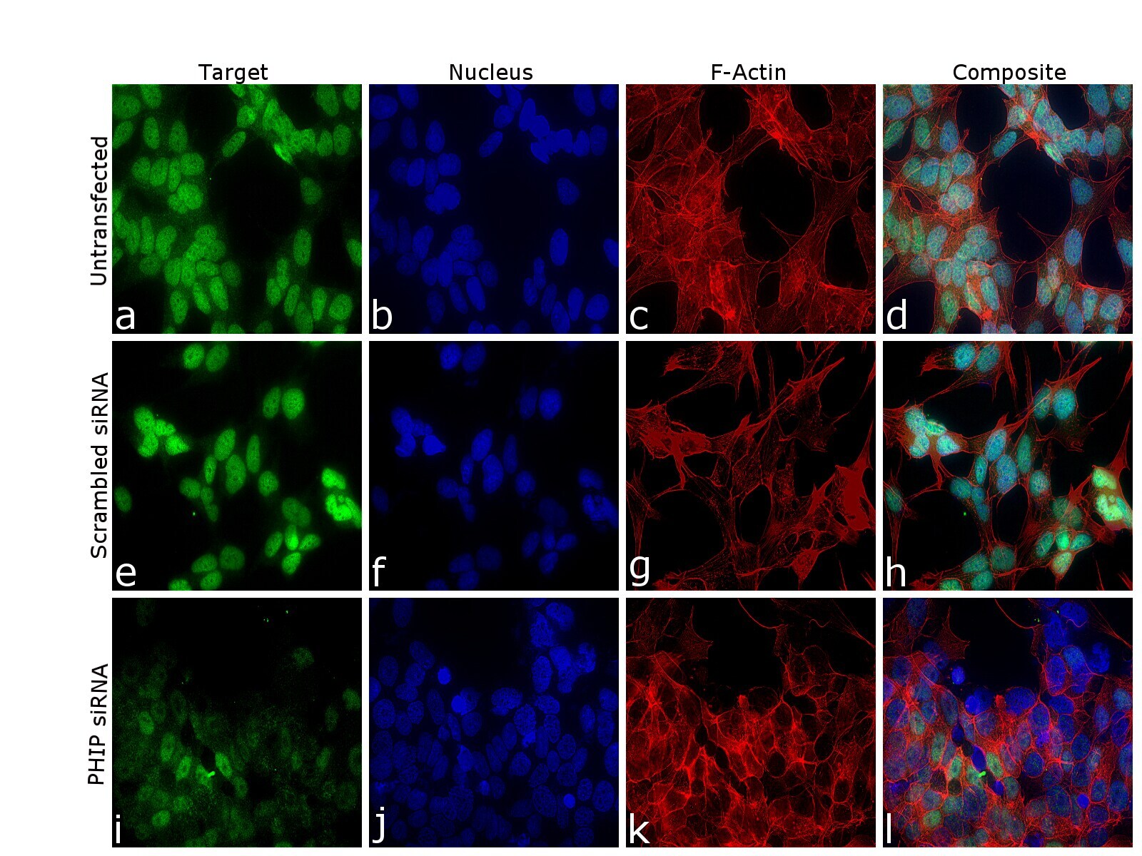
- Knockdown of PH-interacting protein (PHIP) was achieved by transfecting SH-SY5Y cells with PHIP specific siRNA (Silencer® select Product # s30010, s30012). Immunofluorescence analysis was performed on untransfected SH-SY5Y cells (panel a-d), transfected with non-specific scrambled siRNA (panels e-h) and transfected with PHIP specific siRNA (panel i-l). Cells were fixed, permeabilized, and labelled with PHIP Polyclonal Antibody (Product # PA5-77167, 1:100) followed by Goat anti-Rabbit IgG (H+L) Highly Cross-Adsorbed Secondary Antibody, Alexa Fluor Plus 488 (Product # A32731), (1:2500 dilution). Nuclei (blue) were stained using ProLong™ Diamond Antifade Mountant with DAPI (Product # P36962), and Rhodamine Phalloidin (Product # R415, 1:300) was used for cytoskeletal F-actin (Red) staining. Reduction of specific signal was observed upon siRNA mediated knockdown (panel i,l) confirming specificity of the antibody to PHIP (Red). The images were captured at 60X magnification in EVOS™ M7000 Imaging System (Product # AMF7000).