Explore
 Validate
 Learn
Learn Western blot
Antibody data
- Antibody Data
- Antigen structure
- References [0]
- Comments [0]
- Validations
- Western blot [5]
- Immunocytochemistry [1]
- Immunohistochemistry [4]
Submit
Validation data
Reference
Comment
Report error
- Product number
- GTX113954 - Provider product page

- Provider
- GeneTex
- Proper citation
- GeneTex Cat#GTX113954, RRID:AB_11168130
- Product name
- PABP antibody
- Antibody type
- Polyclonal
- Reactivity
- Human, Mouse, Rat
- Host
- Rabbit
No comments: Submit comment
Supportive validation
- Submitted by
- GeneTex (provider)
- Main image
- Experimental details
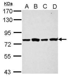
- Sample (30 ?g of whole cell lysate) A: A549 B: H1299 C: HCT116 D: MCF-7 7.5% SDS PAGE GTX113954 diluted at 1:1000 The HRP-conjugated anti-rabbit IgG antibody (GTX213110-01) was used to detect the primary antibody.

- Submitted by
- GeneTex (provider)
- Main image
- Experimental details
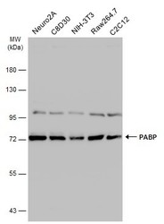
- Various whole cell extracts (30 ?g) were separated by 7.5% SDS-PAGE, and the membrane was blotted with PABP antibody (GTX113954) diluted at 1:1000. The HRP-conjugated anti-rabbit IgG antibody (GTX213110-01) was used to detect the primary antibody.
- Submitted by
- GeneTex (provider)
- Main image
- Experimental details
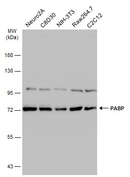
- Various whole cell extracts (30 ?g) were separated by 7.5% SDS-PAGE, and the membrane was blotted with PABP antibody (GTX113954) diluted at 1:1000. The HRP-conjugated anti-rabbit IgG antibody (GTX213110-01) was used to detect the primary antibody.
- Submitted by
- GeneTex (provider)
- Main image
- Experimental details
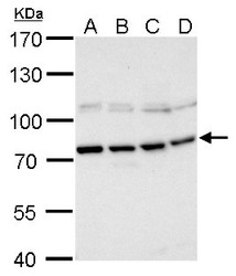
- PABP antibody detects PABP protein by Western blot analysis.A. 30 £gg 293T whole cell lysate/extractB. 30 £gg A431 whole cell lysate/extractC. 30 £gg HeLa whole cell lysate/extractD. 30 £gg HepG2 whole cell lysate/extract7.5 % SDS-PAGEPABP antibody (GTX113954) dilution: 1:1000

- Submitted by
- GeneTex (provider)
- Main image
- Experimental details
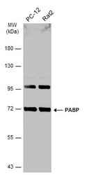
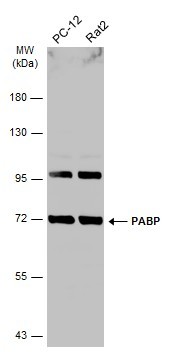
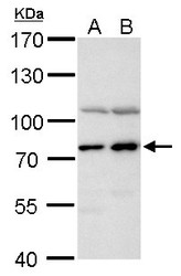
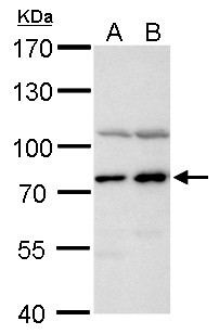
- PABP antibody detects PABP protein by Western blot analysis.A. 30 £gg A549 whole cell lysate/extractB. 30 £gg H1299 whole cell lysate/extract7.5 % SDS-PAGEPABP antibody (GTX113954) dilution: 1:1000
Supportive validation
- Submitted by
- GeneTex (provider)
- Main image
- Experimental details
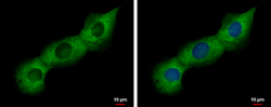
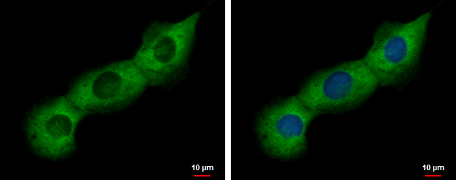
- PABP antibody detects PABP protein at cytoplasm by immunofluorescent analysis.Sample: H1299 cells were fixed in 4% paraformaldehyde at RT for 15 min.Green: PABP protein stained by PABP antibody (GTX113954) diluted at 1:500.Blue: Hoechst 33342 staining.
Supportive validation
- Submitted by
- GeneTex (provider)
- Main image

- Experimental details
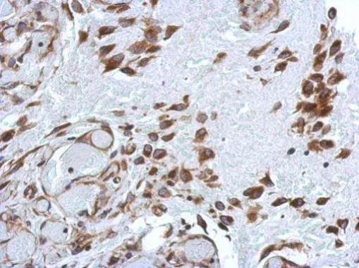
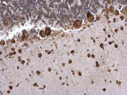
- Immunohistochemical analysis of paraffin-embedded human hepatoma, using PABP(GTX113954) antibody at 1:500 dilution.
- Submitted by
- GeneTex (provider)
- Main image
- Experimental details
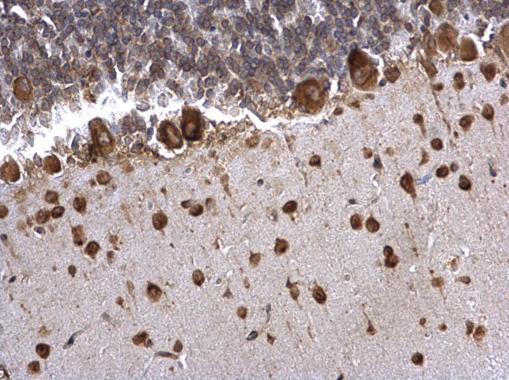
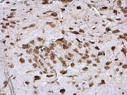
- PABP antibody detects PABP protein at cytosol on mouse middle brain by immunohistochemical analysis. Sample: Paraffin-embedded mouse middle brain. PABP antibody (GTX113954) dilution: 1:500.
- Submitted by
- GeneTex (provider)
- Main image

- Experimental details
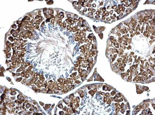
- PABP antibody detects PABP protein at cytosol on mouse testis by immunohistochemical analysis. Sample: Paraffin-embedded mouse testis. PABP antibody (GTX113954) dilution: 1:500.
- Submitted by
- GeneTex (provider)
- Main image
- Experimental details
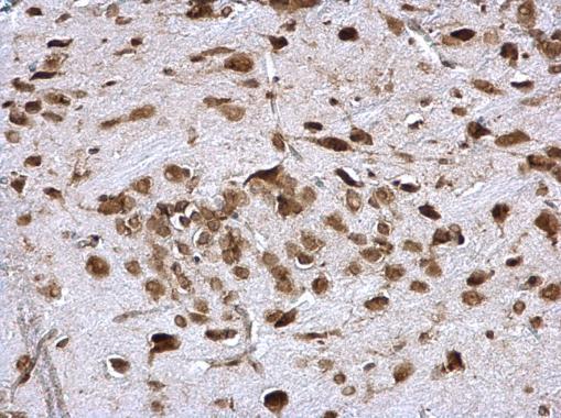
- PABP antibody detects PABP protein at cytosol on rat hind brain by immunohistochemical analysis. Sample: Paraffin-embedded rat hind brain. PABP antibody (GTX113954) dilution: 1:500.