 Explore
 Validate
 Learn
Learn Western blot
Antibody data
- Antibody Data
- Antigen structure
- References [0]
- Comments [0]
- Validations
- Western blot [3]
- Immunocytochemistry [2]
- Other assay [1]
Submit
Validation data
Reference
Comment
Report error
- Product number
- PA5-66900 - Provider product page

- Provider
- Invitrogen Antibodies
- Product name
- PABP Polyclonal Antibody
- Antibody type
- Polyclonal
- Antigen
- Recombinant full-length protein
- Description
- Immunogen sequence: QTQNRAAYYPP SQIAQLRPSP RWTAQG Highest antigen sequence identity to the following orthologs - mouse 100%, rat 100%.
- Reactivity
- Human
- Host
- Rabbit
- Isotype
- IgG
- Vial size
- 100 µL
- Concentration
- 0.2 mg/mL
- Storage
- Store at 4°C short term. For long term storage, store at -20°C, avoiding freeze/thaw cycles.
No comments: Submit comment
Supportive validation
- Submitted by
- Invitrogen Antibodies (provider)
- Main image
- Experimental details
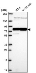
- Western blot analysis of PABP in human cell line RT-4 and human cell line U-251 MG. Samples were probed using a PABP Polyclonal Antibody (Product # PA5-66900).
- Submitted by
- Invitrogen Antibodies (provider)
- Main image

- Experimental details
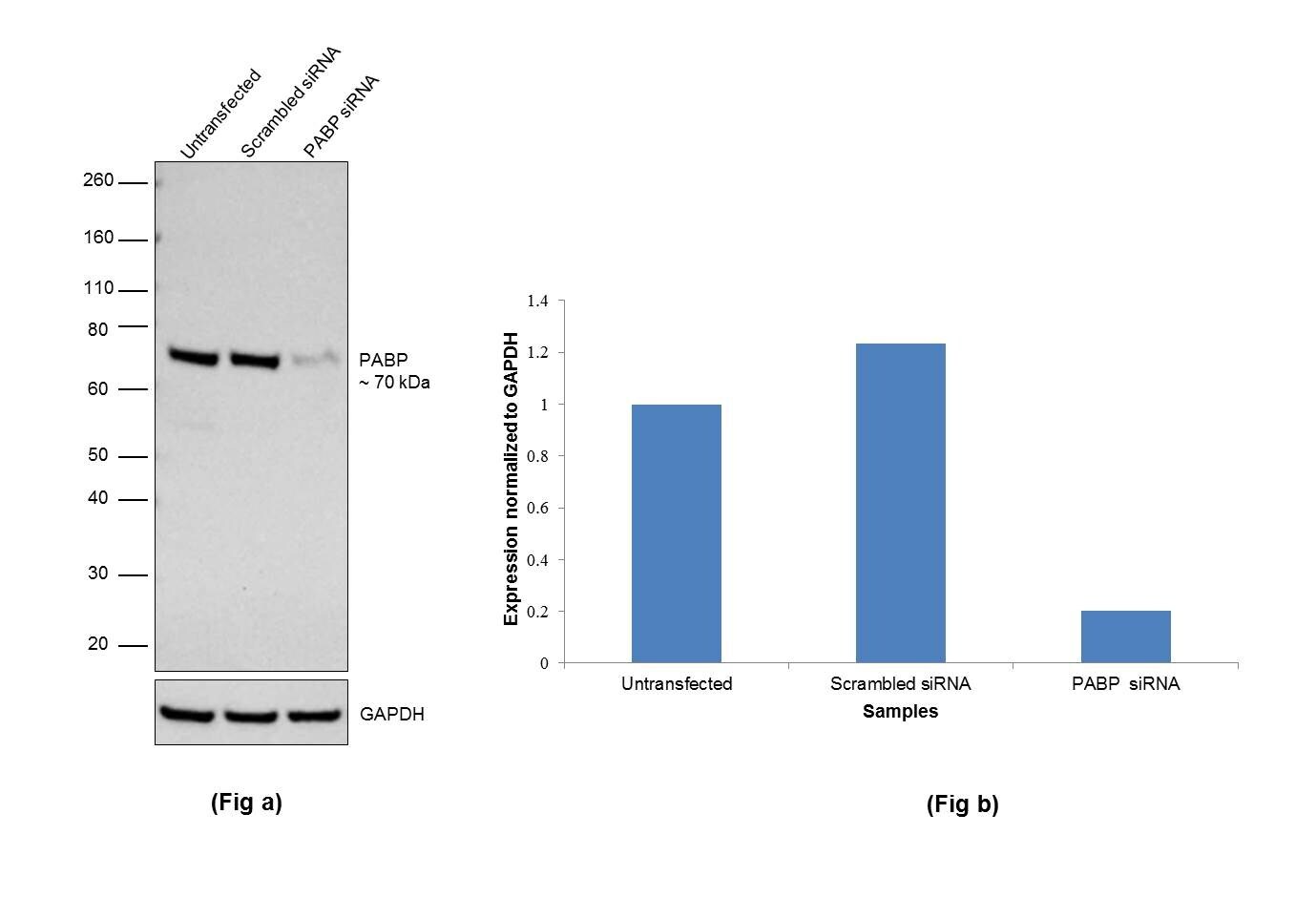
- Knockdown of PABP was achieved by transfecting HeLa cells with PABP specific siRNA (Silencer® select Product # s24290). Western blot analysis (Fig. a) was performed using whole cell extracts from the PABP knockdown cells (lane 3), non-specific scrambled siRNA transfected cells (lane 2) and untransfected cells (lane 1). The blot was probed with PABP Polyclonal Antibody (Product # PA5-66900, 1:500 dilution) and Goat anti-Rabbit IgG (H+L) Superclonal™ Secondary Antibody, HRP conjugate (Product # A27036, 1:4000 dilution). Densitometric analysis of this western blot is shown in histogram (Fig. b). Decrease in signal upon siRNA mediated knock down confirms that antibody is specific to PABP.
- Submitted by
- Invitrogen Antibodies (provider)
- Main image
- Experimental details
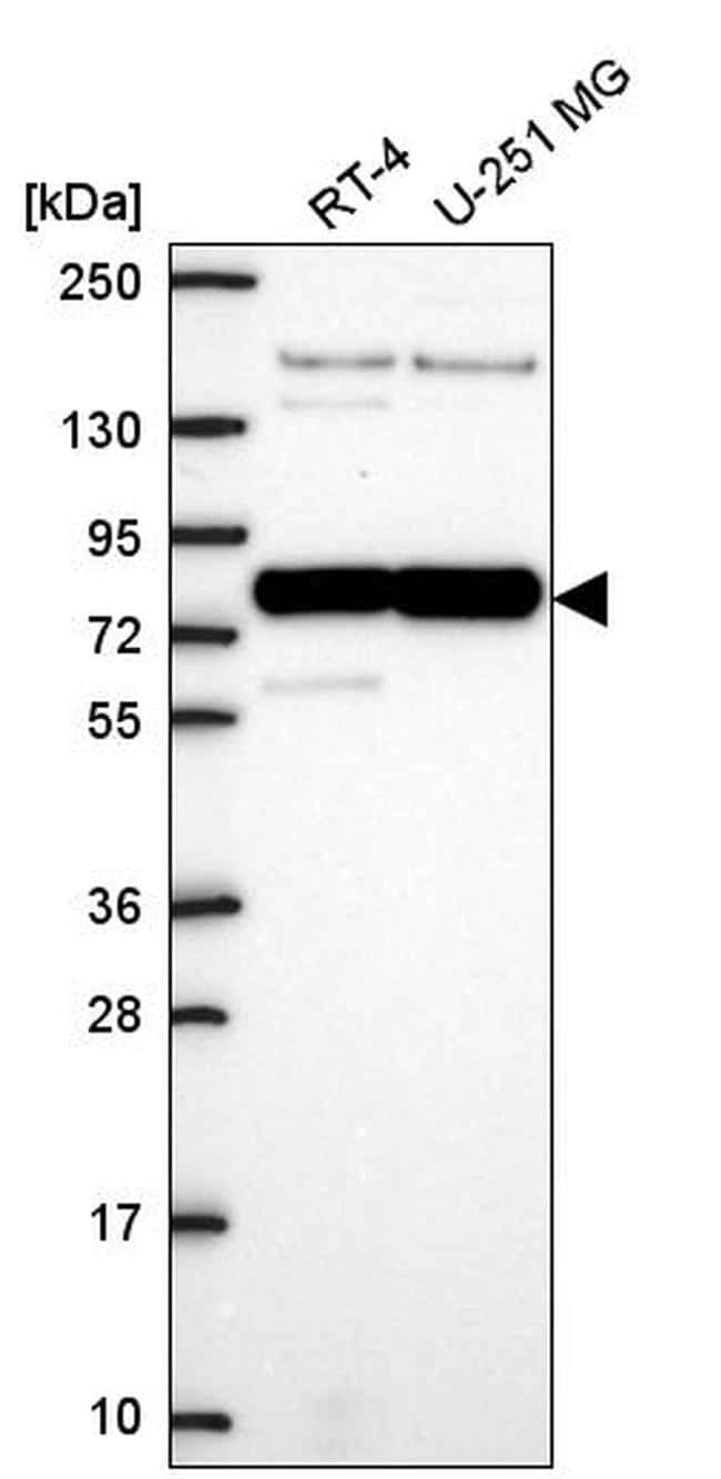
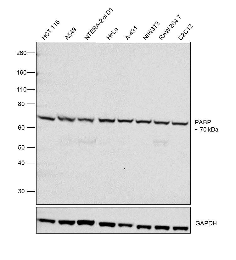
- Western blot was performed using PABP Polyclonal Antibody (Product # PA5-66900) and a 70 kDa band corresponding to PABP was observed across cell lines tested. Whole cell extracts (30 µg lysate) of HCT 116 (Lane 1), A549 (Lane 2), NTERA-2 cl.D1 (Lane 3), HeLa (Lane 4), A-431 (Lane 5), NIH/3T3 (Lane 6), RAW 264.7 (Lane 7) and C2C12 (Lane 8) were electrophoresed using Novex® NuPAGE® 4-12 % Bis-Tris gel (Product # NP0322BOX). Resolved proteins were then transferred onto a nitrocellulose membrane (Product # IB23001) by iBlot® 2 Dry Blotting System (Product # IB21001). The blot was probed with the primary antibody (1:500 dilution) and detected by chemiluminescence using Goat Anti-Rabbit IgG Secondary Antibody, HRP conjugate (Product # A27036, 1:4000 dilution) using the iBright FL 1000 (Product # A32752). Chemiluminescent detection was performed using Novex® ECL Chemiluminescent Substrate Reagent Kit (Product # WP20005).
Supportive validation
- Submitted by
- Invitrogen Antibodies (provider)
- Main image
- Experimental details
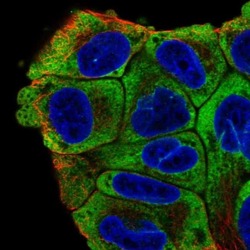
- Immunofluorescent staining of PABP in human cell line RT4 shows localization to cytosol. Samples were probed using a PABP Polyclonal Antibody (Product # PA5-66900).

- Submitted by
- Invitrogen Antibodies (provider)
- Main image
- Experimental details
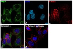
- Immunofluorescence analysis of PABP was performed using 70% confluent log phase HCT 116 cells. The cells were fixed with 4% paraformaldehyde for 10 minutes, permeabilized with 0.1% Triton™ X-100 for 15 minutes, and blocked with 1% BSA for 1 hour at room temperature. The cells were labeled with PABP Rabbit Polyclonal Antibody (Product # PA5-66900) at 4 µg/mL dilution in 0.1% BSA, incubated at 4 degree Celsius overnight and then labeled with Goat anti-Rabbit IgG (H+L) Superclonal™ Secondary Antibody, Alexa Fluor® 488 conjugate (Product # A27034) at a dilution of 1:2000 for 45 minutes at room temperature (Panel a: green). Nuclei (Panel b: blue) were stained with ProLong™ Diamond Antifade Mountant with DAPI (Product # P36962). F-actin (Panel c: red) was stained with Rhodamine Phalloidin (Product # R415). Panel d represents the merged image showing cytoplasmic localization. Panel e represents control cells with no primary antibody to assess background. The images were captured at 60X magnification.

Supportive validation
- Submitted by
- Invitrogen Antibodies (provider)
- Main image
- Experimental details
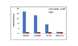
- RNA Immunoprecipitation (RIP) assay of endogenous PABP protein using Anti-PABP Antibody: RIP assay was performed using Anti-PABP Recombinant Rabbit Polyclonal Antibody (Product # PA5-66900, 5 ug) on whole cell lysate from Hep G2 cells. Normal Rabbit IgG was used as a negative IP control. RNA purified by RiboPure™ RNA Purification Kit (Product # AM1924) was analyzed by RT-PCR using the Power SYBR® Green RNA-to-CT™ 1-Step Kit (Product # 4389986) with the primers pairs over RPL30, GAPDH, ACTB mRNA and MALAT1 non-coding RNA. Data is presented as fold enrichment of the antibody signal versus the negative control IgG using the comparative CT method.
