Explore
 Validate
 Learn
Learn Western blot
 Immunohistochemistry
Antibody data
- Antibody Data
- Antigen structure
- References [1]
- Comments [0]
- Validations
- Immunohistochemistry [6]
- Other assay [1]
Submit
Validation data
Reference
Comment
Report error
- Product number
- PA5-114485 - Provider product page

- Provider
- Invitrogen Antibodies
- Product name
- SMURF2 Polyclonal Antibody
- Antibody type
- Polyclonal
- Antigen
- Synthetic peptide
- Description
- Positive controls that may be used include: C2C12 Cell Lysate
- Reactivity
- Human, Mouse, Rat
- Host
- Rabbit
- Isotype
- IgG
- Vial size
- 100 μg
- Concentration
- 1 mg/mL
- Storage
- Store at 4°C short term. For long term storage, store at -20°C, avoiding freeze/thaw cycles.
Submitted references miR-542-3p Attenuates Bone Loss and Marrow Adiposity Following Methotrexate Treatment by Targeting sFRP-1 and Smurf2.
Zhang YL, Liu L, Su YW, Xian CJ
International journal of molecular sciences 2021 Oct 12;22(20)
International journal of molecular sciences 2021 Oct 12;22(20)
No comments: Submit comment
Supportive validation
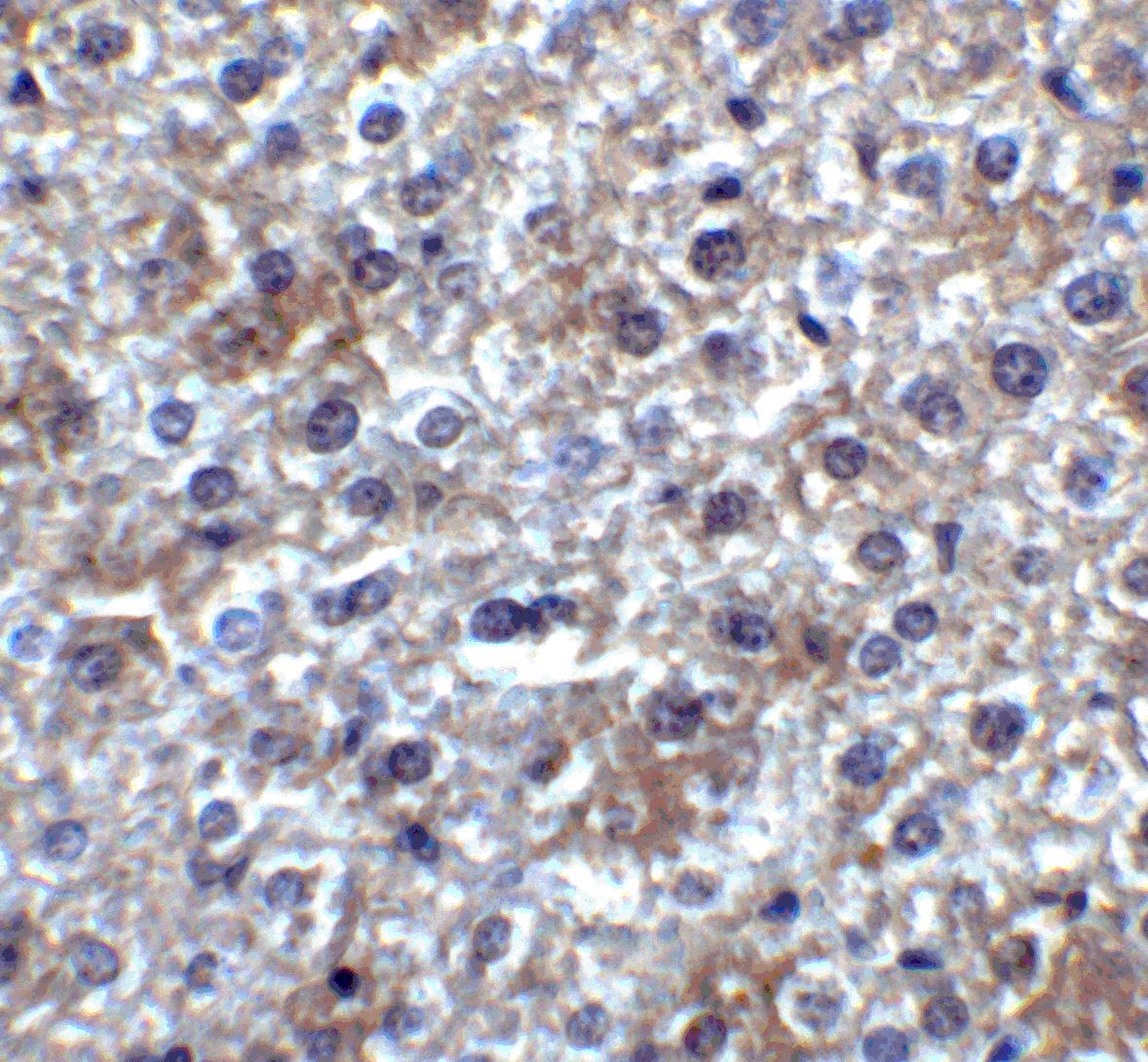
- Submitted by
- Invitrogen Antibodies (provider)
- Main image
- Experimental details
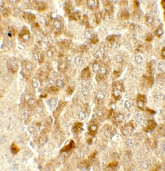
- Immunohistochemistry of SMURF2 in mouse liver tissue with SMURF2 Polyclonal Antibody (Product # PA5-114485) at 2 µg/mL.
- Submitted by
- Invitrogen Antibodies (provider)
- Main image
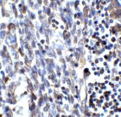
- Experimental details
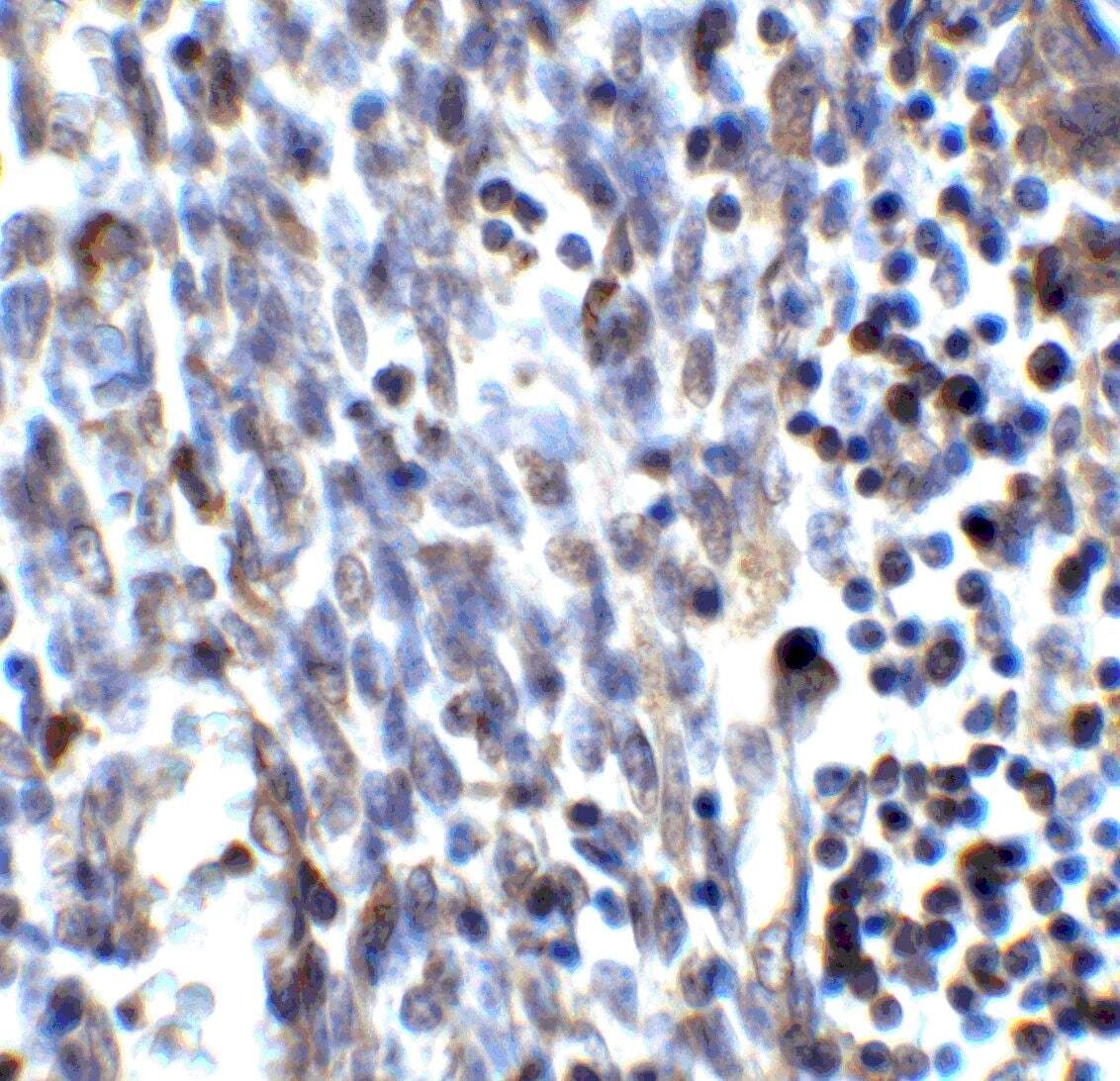
- Immunohistochemistry of SMURF2 in human uterus tissue with SMURF2 Polyclonal Antibody (Product # PA5-114485) at 2 µg/mL.
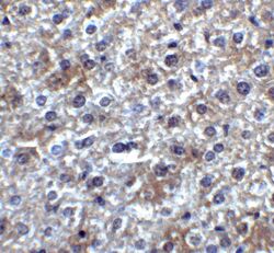
- Submitted by
- Invitrogen Antibodies (provider)
- Main image
- Experimental details
- Immunohistochemistry of SMURF2 in mouse liver tissue with SMURF2 Polyclonal Antibody (Product # PA5-114485) at 5 µg/mL.

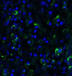
- Submitted by
- Invitrogen Antibodies (provider)
- Main image
- Experimental details
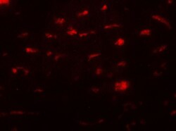
- Immunofluorescence of SMURF2 in mouse liver tissue with SMURF2 Polyclonal Antibody (Product # PA5-114485) at 20 µg/mL.
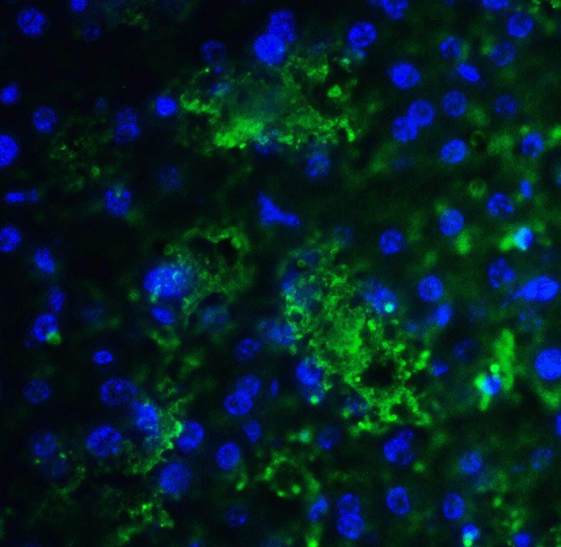
- Submitted by
- Invitrogen Antibodies (provider)
- Main image

- Experimental details
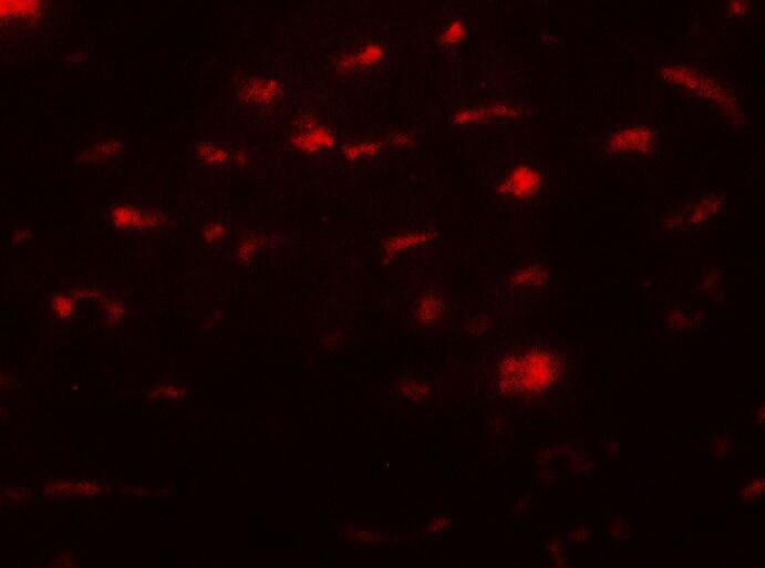
- Immunofluorescence of SMURF2 in mouse liver tissue with SMURF2 Polyclonal Antibody (Product # PA5-114485) at 20 µg/mL. Green: SMURF2 Blue: DAPI staining
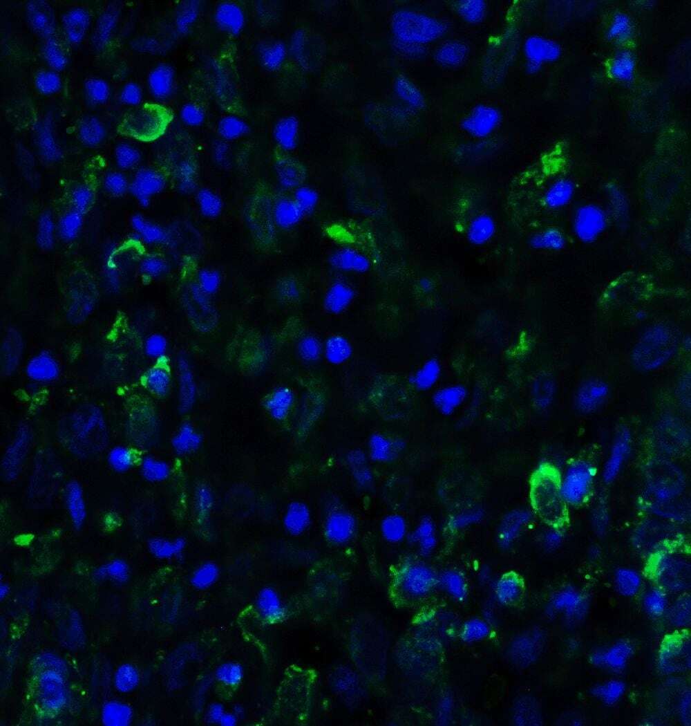
- Submitted by
- Invitrogen Antibodies (provider)
- Main image
- Experimental details
- Immunofluorescence of SMURF2 in human uterus tissue with SMURF2 Polyclonal Antibody (Product # PA5-114485) at 20 µg/mL. Green: SMURF2 Blue: DAPI staining
Supportive validation
- Submitted by
- Invitrogen Antibodies (provider)
- Main image

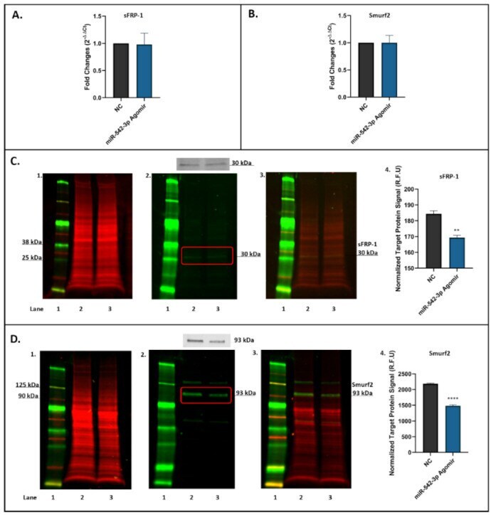
- Experimental details
- Figure 4 miR-542-3p Inhibits sFRP-1 and Smurf2 expression at the post-transcriptional level in MC3T3.E1 osteoblastic cells. ( A ) RT-qPCR results showed that there were no significant changes in sFRP-1 mRNA expression after miRNA-542-3p transfection when compared to the negative control (NC) group. ( B ) RT-qPCR analyses of Smurf2 mRNA expression after miRNA-542-3p transfection, which was not significantly changed compared to the NC. ( C ) Western blot studies of sFPR-1 after miRNA-542-3p transfection. The protein expression levels of sFRP-1 (~30 kDa) in treated cells were notably decreased compared with NC. ( C1 ). Total protein extract was visualized (700 nm channel, red); ( C2 ). Target protein sFRP-1 was visualized (800 nm channel, green); ( C3 ). Merged images of blots; and ( C4 ). Normalized target sFRP-1 protein signal (from 3 experiments). ( D ) Western blot studies of Smurf2 after miRNA-542-3p transfection. The protein expression levels of Smurf2 (~93 kDa) in treated cells were notably decreased compared with NC. ( D1 ). Total protein extract was visualized (700 nm channel, red); ( D2 ). Target protein Smurf2 was visualized (800 nm channel, green); ( D3 ). Merged images of blots; and ( D4 ). Normalized target Smurf2 protein signal (from 3 experiments). Treatments on each lane: lane 1: pre-stained protein ladder; lane 2: negative control (NC); lane 3: miR-542-3p agomir. R.F.U: relative fluorescence units. Statistical significance analyses were performed via t test. Si