Explore
 Validate
 Learn
Learn Western blot
 Immunocytochemistry
Antibody data
- Antibody Data
- Antigen structure
- References [5]
- Comments [0]
- Validations
- Immunocytochemistry [4]
- Immunohistochemistry [10]
- Other assay [5]
Submit
Validation data
Reference
Comment
Report error
- Product number
- PA5-29326 - Provider product page

- Provider
- Invitrogen Antibodies
- Product name
- HK2 Polyclonal Antibody
- Antibody type
- Polyclonal
- Antigen
- Recombinant full-length protein
- Description
- Recommended positive controls: U87-MG, SK-N-SH, IMR32, SK-N-AS, Mouse brain. Predicted reactivity: Mouse (97%), Rat (97%), Zebrafish (86%), Pig (96%), Chicken (88%). Store product as a concentrated solution. Centrifuge briefly prior to opening the vial.
- Reactivity
- Human, Mouse, Rat
- Host
- Rabbit
- Isotype
- IgG
- Vial size
- 100 μL
- Concentration
- 0.63 mg/mL
- Storage
- Store at 4°C short term. For long term storage, store at -20°C, avoiding freeze/thaw cycles.
Submitted references circATP2A2 promotes osteosarcoma progression by upregulating MYH9.
Tanshinone IIA inhibits glucose metabolism leading to apoptosis in cervical cancer.
BACH1 Stabilization by Antioxidants Stimulates Lung Cancer Metastasis.
Cyclopamine tartrate, a modulator of hedgehog signaling and mitochondrial respiration, effectively arrests lung tumor growth and progression.
Hexokinase 2 drives glycogen accumulation in equine endometrium at day 12 of diestrus and pregnancy.
Cao X, Meng X, Fu P, Wu L, Yang Z, Chen H
Open medicine (Warsaw, Poland) 2021;16(1):1749-1761
Open medicine (Warsaw, Poland) 2021;16(1):1749-1761
Tanshinone IIA inhibits glucose metabolism leading to apoptosis in cervical cancer.
Liu Z, Zhu W, Kong X, Chen X, Sun X, Zhang W, Zhang R
Oncology reports 2019 Nov;42(5):1893-1903
Oncology reports 2019 Nov;42(5):1893-1903
BACH1 Stabilization by Antioxidants Stimulates Lung Cancer Metastasis.
Wiel C, Le Gal K, Ibrahim MX, Jahangir CA, Kashif M, Yao H, Ziegler DV, Xu X, Ghosh T, Mondal T, Kanduri C, Lindahl P, Sayin VI, Bergo MO
Cell 2019 Jul 11;178(2):330-345.e22
Cell 2019 Jul 11;178(2):330-345.e22
Cyclopamine tartrate, a modulator of hedgehog signaling and mitochondrial respiration, effectively arrests lung tumor growth and progression.
Kalainayakan SP, Ghosh P, Dey S, Fitzgerald KE, Sohoni S, Konduri PC, Garrossian M, Liu L, Zhang L
Scientific reports 2019 Feb 5;9(1):1405
Scientific reports 2019 Feb 5;9(1):1405
Hexokinase 2 drives glycogen accumulation in equine endometrium at day 12 of diestrus and pregnancy.
Bramer SA, Macedo A, Klein C
Reproductive biology and endocrinology : RB&E 2017 Jan 5;15(1):4
Reproductive biology and endocrinology : RB&E 2017 Jan 5;15(1):4
No comments: Submit comment
Supportive validation
- Submitted by
- Invitrogen Antibodies (provider)
- Main image
- Experimental details
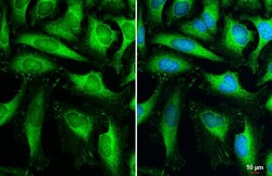
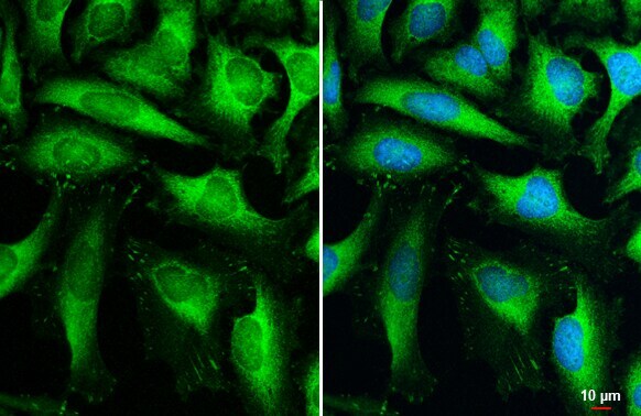
- Immunocytochemistry-Immunofluorescence analysis of HK2 was performed in HeLa cells fixed in 4% paraformaldehyde at RT for 15 min. Green: HK2 Polyclonal Antibody (Product # PA5-29326) diluted at 1:2000. Blue: Hoechst 33342 staining. Scale bar = 10 µm.

- Submitted by
- Invitrogen Antibodies (provider)
- Main image
- Experimental details
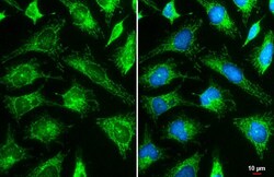
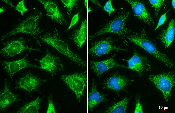
- HK2 Polyclonal Antibody detects Hexokinase II protein at mitochondria by immunofluorescent analysis. Sample: HeLa cells were fixed in ice-cold MeOH for 5 min. Green: Hexokinase II stained by HK2 Polyclonal Antibody (Product # PA5-29326) diluted at 1:2,000. Blue: Fluoroshield with DAPI . Scale bar= 10 µm.
- Submitted by
- Invitrogen Antibodies (provider)
- Main image

- Experimental details
- Immunocytochemistry-Immunofluorescence analysis of HK2 was performed in HeLa cells fixed in 4% paraformaldehyde at RT for 15 min. Green: HK2 Polyclonal Antibody (Product # PA5-29326) diluted at 1:2000. Blue: Hoechst 33342 staining. Scale bar = 10 µm.
- Submitted by
- Invitrogen Antibodies (provider)
- Main image

- Experimental details
- HK2 Polyclonal Antibody detects Hexokinase II protein at mitochondria by immunofluorescent analysis. Sample: HeLa cells were fixed in ice-cold MeOH for 5 min. Green: Hexokinase II stained by HK2 Polyclonal Antibody (Product # PA5-29326) diluted at 1:2,000. Blue: Fluoroshield with DAPI . Scale bar= 10 µm.

Supportive validation
- Submitted by
- Invitrogen Antibodies (provider)
- Main image
- Experimental details
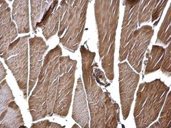
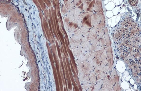
- HK2 Polyclonal Antibody detects Hexokinase II protein at cytosol on mouse muscle by immunohistochemical analysis. Sample: Paraffin-embedded mouse muscle. HK2 Polyclonal Antibody (Product # PA5-29326) dilution: 1:500. Antigen Retrieval: EDTA based buffer, pH 8.0, 15 min.
- Submitted by
- Invitrogen Antibodies (provider)
- Main image
- Experimental details
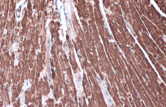
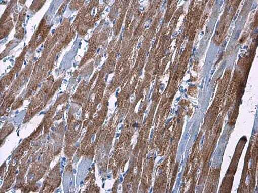
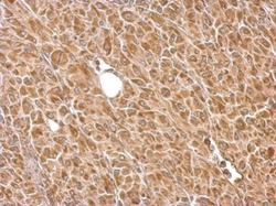
- HK2 Polyclonal Antibody detects Hexokinase II protein at cytoplasm by immunohistochemical analysis. Sample: Paraffin-embedded mouse heart. Hexokinase II stained by HK2 Polyclonal Antibody (Product # PA5-29326) diluted at 1:2,000. Antigen Retrieval: Citrate buffer, pH 6.0, 15 min.
- Submitted by
- Invitrogen Antibodies (provider)
- Main image
- Experimental details
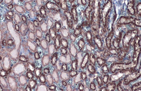
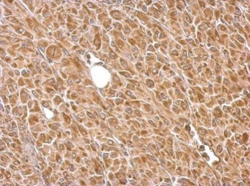
- HK2 Polyclonal Antibody detects Hexokinase II protein at cytoplasm by immunohistochemical analysis. Sample: Paraffin-embedded rat kidney. Hexokinase II stained by HK2 Polyclonal Antibody (Product # PA5-29326) diluted at 1:2,000. Antigen Retrieval: Citrate buffer, pH 6.0, 15 min.
- Submitted by
- Invitrogen Antibodies (provider)
- Main image

- Experimental details
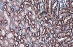
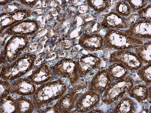
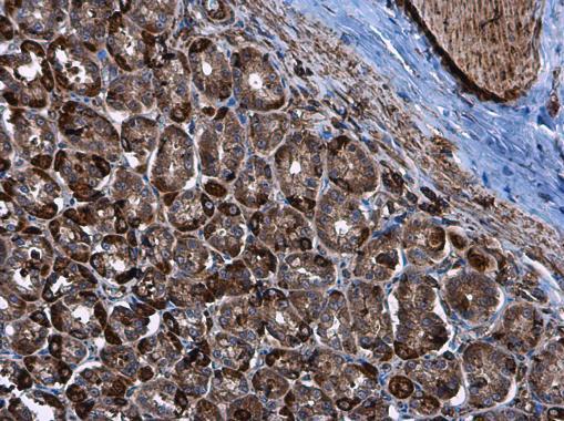
- Immunohistochemistry (Paraffin) analysis of HK2 was performed in paraffin-embedded rat kidney tissue using HK2 Polyclonal Antibody (Product # PA5-29326) at a dilution of 1:500.
- Submitted by
- Invitrogen Antibodies (provider)
- Main image

- Experimental details
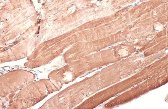
- Immunohistochemistry (Paraffin) analysis of HK2 was performed in paraffin-embedded rat heart tissue using HK2 Polyclonal Antibody (Product # PA5-29326) at a dilution of 1:500.
- Submitted by
- Invitrogen Antibodies (provider)
- Main image
- Experimental details
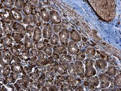
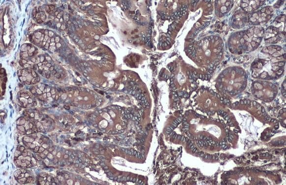
- Immunohistochemistry (Paraffin) analysis of HK2 was performed in paraffin-embedded rat stomach tissue using HK2 Polyclonal Antibody (Product # PA5-29326) at a dilution of 1:500.
- Submitted by
- Invitrogen Antibodies (provider)
- Main image

- Experimental details
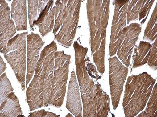
- HK2 Polyclonal Antibody detects Hexokinase II protein at cytoplasm by immunohistochemical analysis. Sample: Paraffin-embedded mouse muscle. Hexokinase II stained by HK2 Polyclonal Antibody (Product # PA5-29326) diluted at 1:500. Antigen Retrieval: Citrate buffer, pH 6.0, 15 min.
- Submitted by
- Invitrogen Antibodies (provider)
- Main image

- Experimental details
- HK2 Polyclonal Antibody detects Hexokinase II protein at cytoplasm by immunohistochemical analysis. Sample: Paraffin-embedded rat colon. Hexokinase II stained by HK2 Polyclonal Antibody (Product # PA5-29326) diluted at 1:2,000. Antigen Retrieval: Citrate buffer, pH 6.0, 15 min.
- Submitted by
- Invitrogen Antibodies (provider)
- Main image

- Experimental details
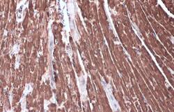
- HK2 Polyclonal Antibody detects Hexokinase II protein at cytoplasm by immunohistochemical analysis. Sample: Paraffin-embedded rat muscle. Hexokinase II stained by HK2 Polyclonal Antibody (Product # PA5-29326) diluted at 1:1,000. Antigen Retrieval: Citrate buffer, pH 6.0, 15 min.
- Submitted by
- Invitrogen Antibodies (provider)
- Main image
- Experimental details
- HK2 Polyclonal Antibody detects HK2 protein at cytosol on U87 xenograft by immunohistochemical analysis. Sample: Paraffin-embedded U87 xenograft. HK2 Polyclonal Antibody (Product # PA5-29326) dilution: 1:500. Antigen Retrieval: EDTA based buffer, pH 8.0, 15 min.
Supportive validation
- Submitted by
- Invitrogen Antibodies (provider)
- Main image
- Experimental details
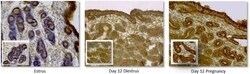
- Fig. 2 Immunohistochemical analysis of HK2 protein distribution in equine endometrium at estrus, and Day 12 of diestrus and pregnancy. Small inserted panels show glands located in the stratum compactum. No staining was observed in the negative control (not shown)

- Submitted by
- Invitrogen Antibodies (provider)
- Main image

- Experimental details
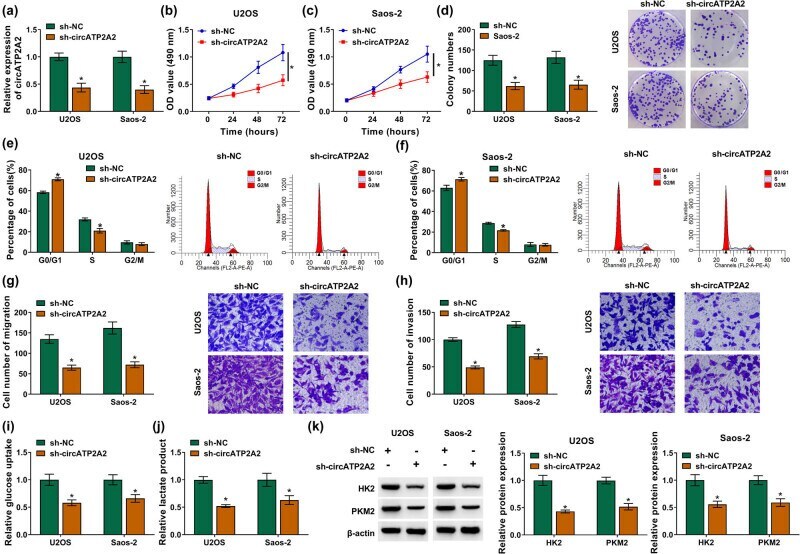
- Figure 2 circATP2A2 promoted OS cell malignancy and glycolysis. (a-k) OS cells were transfected with sh-NC or sh-circATP2A2. (a) The interference efficiency of sh-circATP2A2 in OS cells was verified by RT-qPCR. (b-h) The proliferation, cell cycle progression, migration, and invasion of OS cells were evaluated by MTT, plate clone, flow cytometry, and transwell assays, respectively. (i and j) Assessment of glucose uptake and lactate product levels in OS cells. (k) Detection of protein levels of HK2 and PKM2 in OS cells by western blotting. * P < 0.05.
- Submitted by
- Invitrogen Antibodies (provider)
- Main image

- Experimental details
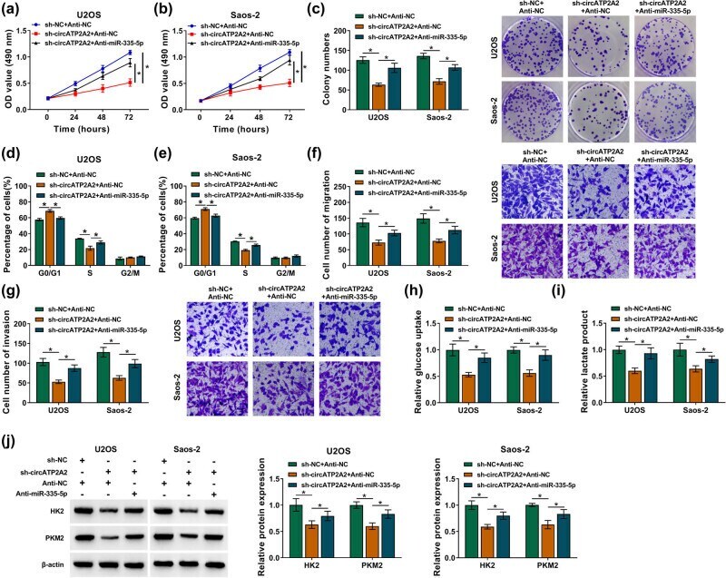
- Figure 4 circATP2A2 regulated OS cell malignancy and glycolysis by sponging miR-335-5p. (a-j) OS cells were transfected with sh-NC + Anti-NC, sh-circATP2A2 + Anti-NC, or sh-circATP2A2 + anti-miR-335-5p. (a-g) The proliferation, cell cycle progression, migration, and invasion of OS cells were determined by MTT, plate clone, flow cytometry, and transwell assays, respectively. (h and i) Analysis of glucose uptake and lactate product levels in OS cells. (j) Protein levels of HK2 and PKM2 in OS cells were measured by Western blotting. * P < 0.05.
- Submitted by
- Invitrogen Antibodies (provider)
- Main image
- Experimental details
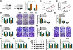
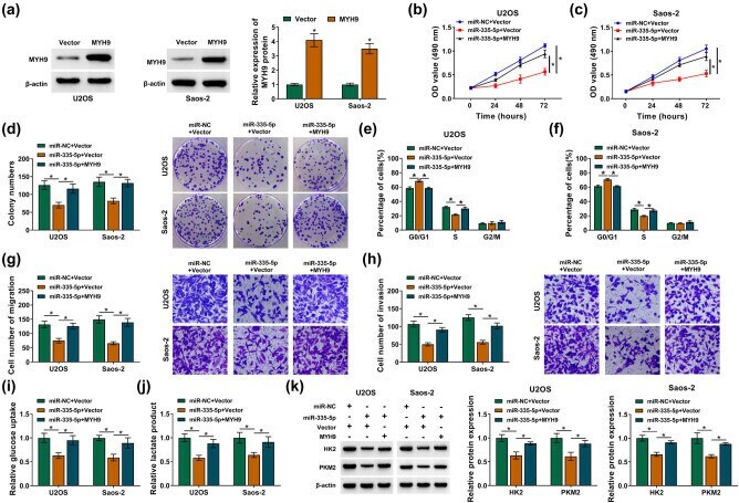
- Figure 6 MiR-335-5p targeted MYH9 to inhibit OS cell malignancy and glycolysis. (a) The overexpression efficiency of MYH9 in OS cells was detected by Western blotting. (b-k) OS cells were transfected with miR-NC + vector, miR-335-5p + vector, or miR-335-5p + MYH9. (b-h) The proliferation, cell cycle progression, migration, and invasion of OS cells were evaluated by MTT, plate clone, flow cytometry, and transwell assays, respectively. (i and j) Detection of glucose uptake and lactate product levels in OS cells. (k) Analysis of HK2 and PKM2 protein levels in U2OS and Saos-2 cells by Western blotting. * P < 0.05.
- Submitted by
- Invitrogen Antibodies (provider)
- Main image
- Experimental details
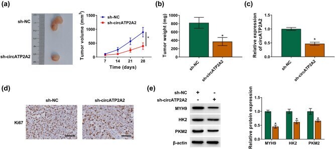
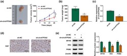
- Figure 7 circATP2A2 inhibition decreased OS cell proliferation and glycolysis in vivo . (a and b) Tumor volume and weight of mice injected with OS cells carrying sh-circATP2A2 or sh-NC. (c) circATP2A2 expression in xenograft tumors in the sh-circATP2A2 and sh-NC groups was analyzed by RT-qPCR. (d) The level of Ki67 in xenograft tumors in the sh-circATP2A2 and sh-NC groups was analyzed by IHC. (e) Protein levels of MYH9, HK2 and PKM2 in xenograft tumors in the sh-circATP2A2 and sh-NC groups were detected by Western blotting. * P < 0.05.