 Explore
 Validate
 Learn
Learn Western blot
 Immunocytochemistry
Antibody data
- Antibody Data
- Antigen structure
- References [0]
- Comments [0]
- Validations
- Immunocytochemistry [2]
- Immunoprecipitation [1]
- Immunohistochemistry [10]
- Other assay [1]
Submit
Validation data
Reference
Comment
Report error
- Product number
- PA5-116079 - Provider product page

- Provider
- Invitrogen Antibodies
- Product name
- TSC1 Polyclonal Antibody
- Antibody type
- Polyclonal
- Antigen
- Synthetic peptide
- Description
- Antibody detects endogenous levels of total TSC1.
- Reactivity
- Human, Mouse, Rat
- Host
- Rabbit
- Isotype
- IgG
- Vial size
- 100 μL
- Concentration
- 1 mg/mL
- Storage
- -20°C
No comments: Submit comment
Supportive validation
- Submitted by
- Invitrogen Antibodies (provider)
- Main image

- Experimental details
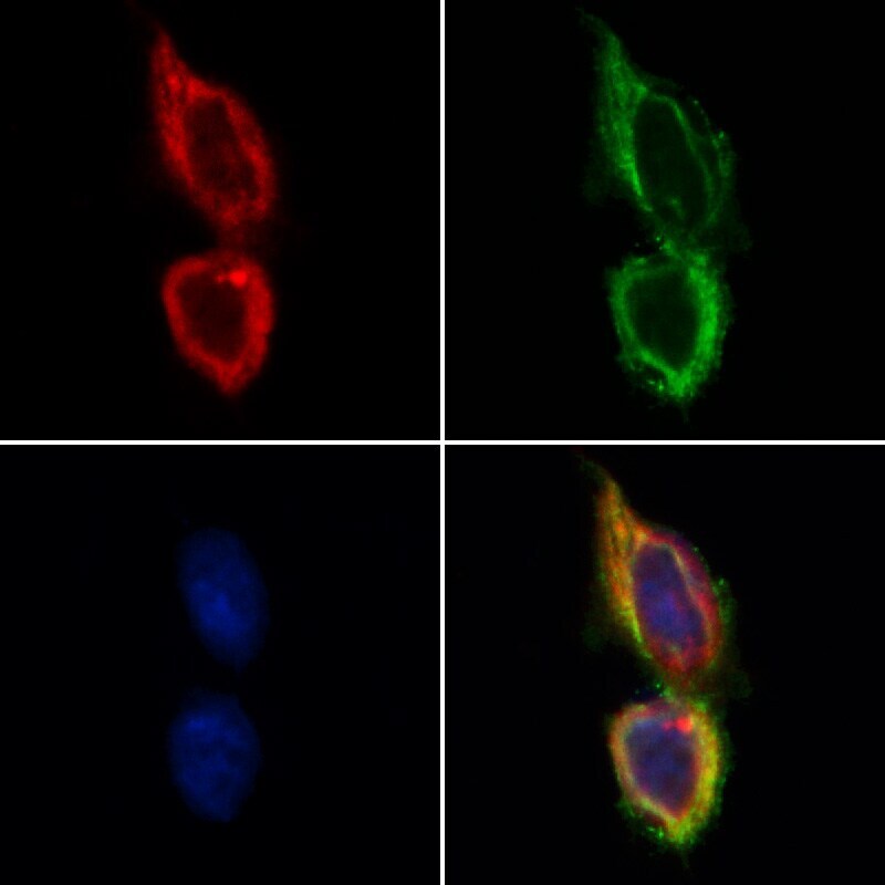
- Immunocytochemistry analysis of TSC1 in HeLa cells. Samples were treated with PFA, permeabilized in 0.1% Triton X-100, blocked in 10% serum (45 min at 25°C), and incubated with polyclonal antibody (Product # PA5-116079) at a dilution of 1:200 (1 hr at 37°C). Secondary staining was applied with mouse anti-beta tubulin; AlexaFluor 594 conjugated goat anti-rabbit IgG (Red); AlexaFluor 88 conjugated goat anti-mouse IgG (Green) and DAPI (blue) using a dilution of 1:200 (1 hr at 37°C).
- Submitted by
- Invitrogen Antibodies (provider)
- Main image

- Experimental details
- Immunocytochemistry analysis of TSC1 in HeLa cells. Samples were treated with PFA, permeabilized in 0.1% Triton X-100, blocked in 10% serum (45 min at 25°C), and incubated with polyclonal antibody (Product # PA5-116079) at a dilution of 1:200 (1 hr at 37°C). Secondary staining was applied with mouse anti-beta tubulin; AlexaFluor 594 conjugated goat anti-rabbit IgG (Red); AlexaFluor 88 conjugated goat anti-mouse IgG (Green) and DAPI (blue) using a dilution of 1:200 (1 hr at 37°C).

Supportive validation
- Submitted by
- Invitrogen Antibodies (provider)
- Main image
- Experimental details
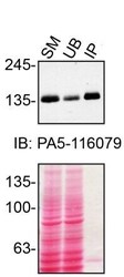
- Immunoprecipitation of TSC1 was performed on HAP1 cell lysates. Antibody-bead conjugates were prepared by adding 2 µg of TSC1 polyclonal antibody (Product # PA5-116079) with 30 µL of protein A-Sepharose beads and rocked overnight at 4°C. 1 mg of lysate was incubated with an antibody-bead conjugate for 2 hours at 4°C. Following centrifugation and multiple washes, 10% starting material (SM), 10% unbound fraction (UB) and immunoprecipitated fraction (IP) were processed for immunoblot using the same antibody. Ponceau stained transfer of blot is shown. Data courtesy of YCharOS Inc., an open science company with the mission of characterizing commercially available antibodies using knockout validation.
Supportive validation
- Submitted by
- Invitrogen Antibodies (provider)
- Main image
- Experimental details
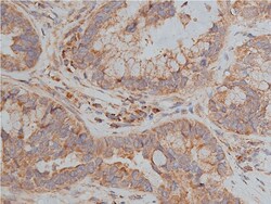
- Immunohistochemistry analysis of TSC1 in human lung cancer tissue sections. Samples were treated with formaldehyde and treated with citrate buffer for antigen retrieval, blocked, and incubated (1.5 hours at 22°C) with polyclonal antibody (Product # PA5-116079) at a dilution of 1:200. Secondary staining was applied with HRP conjugated goat anti-rabbit.

- Submitted by
- Invitrogen Antibodies (provider)
- Main image
- Experimental details
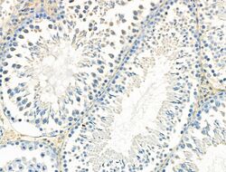
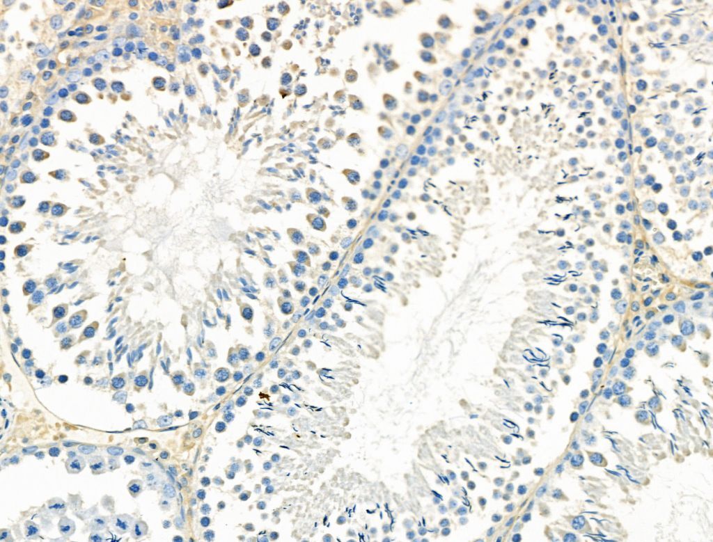
- Immunohistochemistry analysis of TSC1 in rat testis tissue. Samples were treated with formaldehyde and treated with citrate buffer for antigen retrieval, blocked, and incubated (1.5 hours at 22°C) with polyclonal antibody (Product # PA5-116079) at a dilution of 1:100. Secondary staining was applied with HRP conjugated anti-Rabbit.
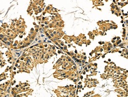
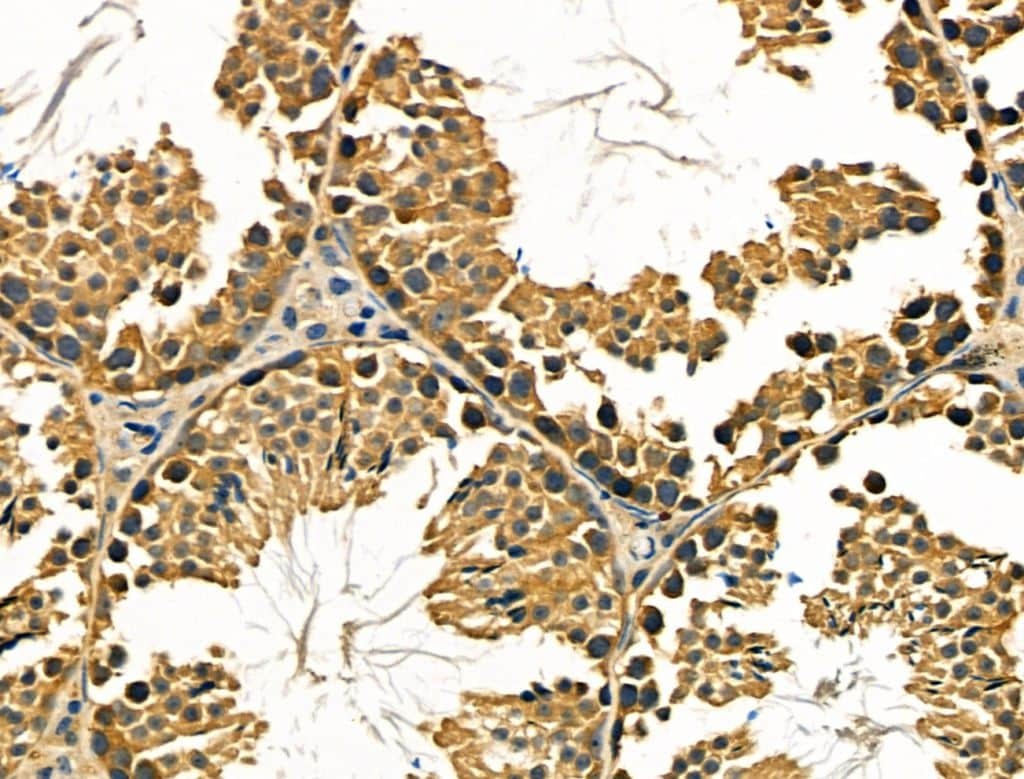
- Submitted by
- Invitrogen Antibodies (provider)
- Main image

- Experimental details
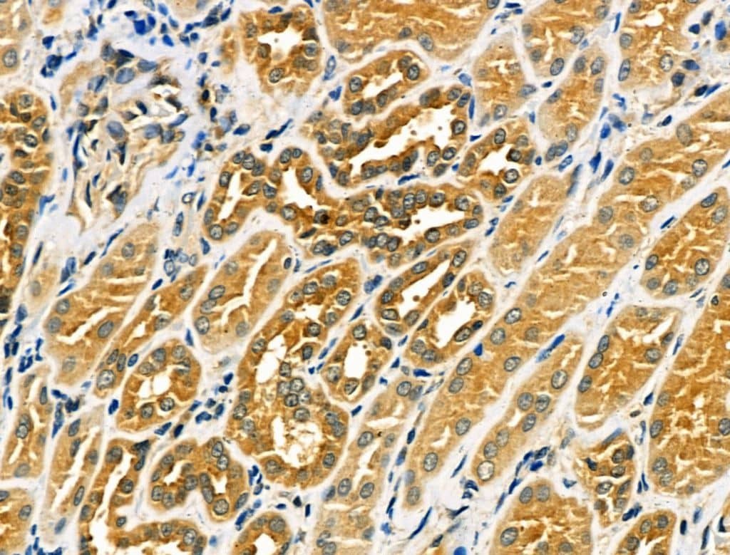
- Immunohistochemistry analysis of TSC1 in human kidney cancer and adjacent normal tissues. Samples were treated with formaldehyde and treated with citrate buffer for antigen retrieval, blocked, and incubated (1.5 hours at 22°C) with polyclonal antibody (Product # PA5-116079) at a dilution of 1:100. Secondary staining was applied with HRP conjugated anti-Rabbit.
- Submitted by
- Invitrogen Antibodies (provider)
- Main image
- Experimental details
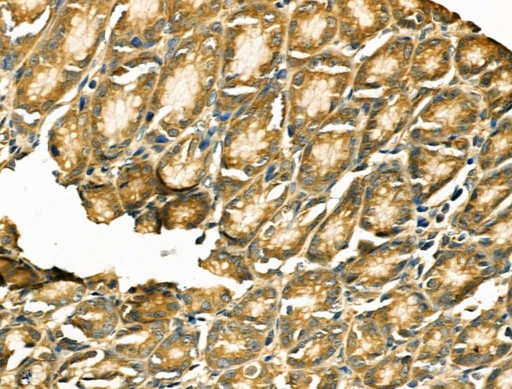
- Immunohistochemistry analysis of TSC1 in mouse kidney tissue. Samples were treated with formaldehyde and treated with citrate buffer for antigen retrieval, blocked, and incubated (1.5 hours at 22°C) with polyclonal antibody (Product # PA5-116079) at a dilution of 1:100. Secondary staining was applied with HRP conjugated anti-Rabbit.

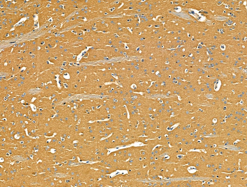
- Submitted by
- Invitrogen Antibodies (provider)
- Main image
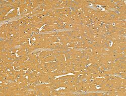
- Experimental details
- Immunohistochemistry analysis of TSC1 in mouse brain tissue. Samples were treated with formaldehyde and treated with citrate buffer for antigen retrieval, blocked, and incubated (1.5 hours at 22°C) with polyclonal antibody (Product # PA5-116079) at a dilution of 1:100. Secondary staining was applied with HRP conjugated anti-Rabbit.
- Submitted by
- Invitrogen Antibodies (provider)
- Main image
- Experimental details
- Immunohistochemistry analysis of TSC1 in mouse testis tissue. Samples were treated with formaldehyde and treated with citrate buffer for antigen retrieval, blocked, and incubated (1.5 hours at 22°C) with polyclonal antibody (Product # PA5-116079) at a dilution of 1:100. Secondary staining was applied with HRP conjugated anti-Rabbit.
- Submitted by
- Invitrogen Antibodies (provider)
- Main image
- Experimental details
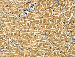
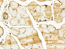
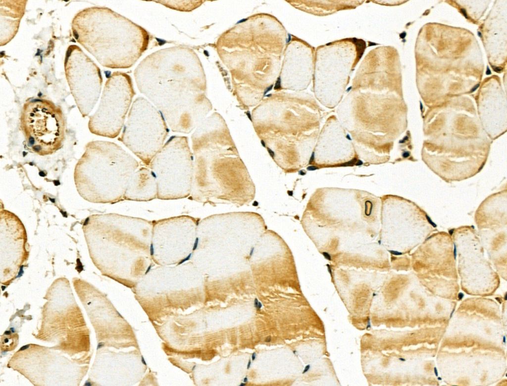
- Immunohistochemistry analysis of TSC1 in mouse muscle tissue. Samples were treated with formaldehyde and treated with citrate buffer for antigen retrieval, blocked, and incubated (1.5 hours at 22°C) with polyclonal antibody (Product # PA5-116079) at a dilution of 1:100. Secondary staining was applied with HRP conjugated anti-Rabbit.
- Submitted by
- Invitrogen Antibodies (provider)
- Main image

- Experimental details
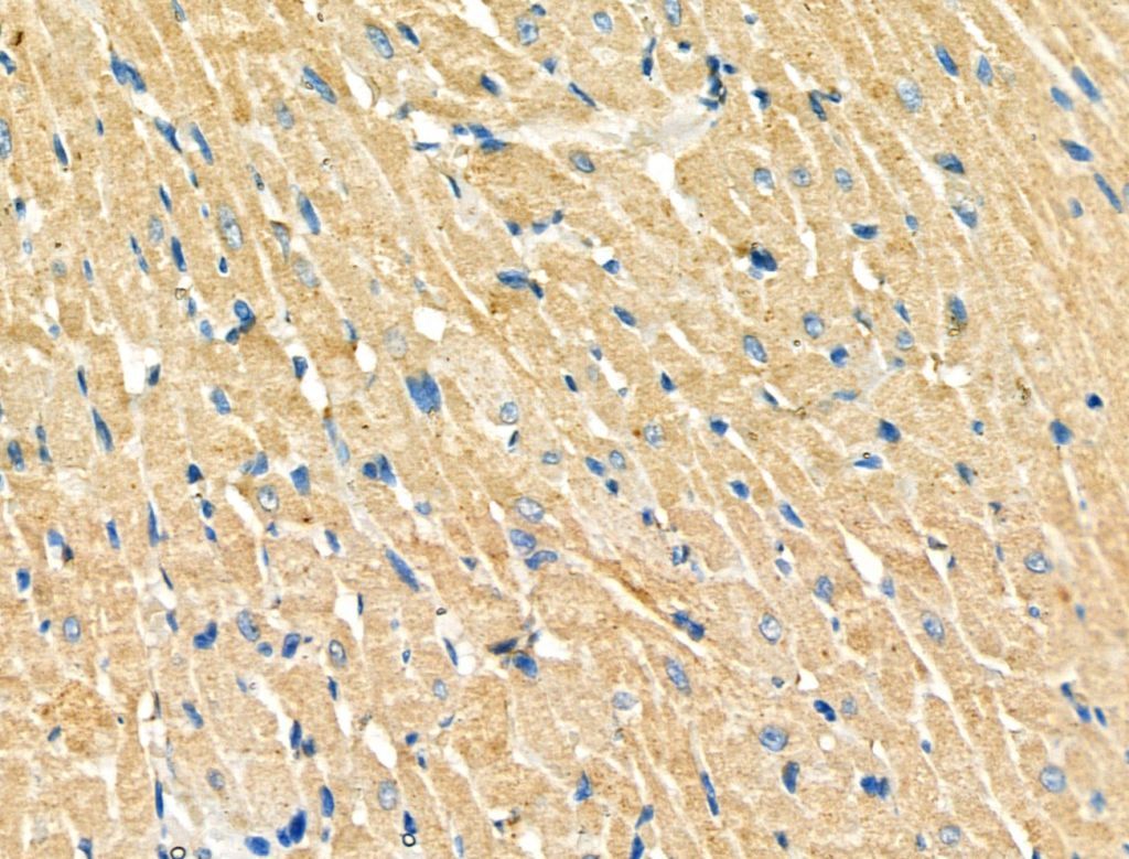
- Immunohistochemistry analysis of TSC1 in rat heart tissue. Samples were treated with formaldehyde and treated with citrate buffer for antigen retrieval, blocked, and incubated (1.5 hours at 22°C) with polyclonal antibody (Product # PA5-116079) at a dilution of 1:100. Secondary staining was applied with HRP conjugated anti-Rabbit.
- Submitted by
- Invitrogen Antibodies (provider)
- Main image
- Experimental details
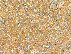
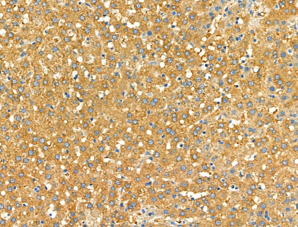
- Immunohistochemistry analysis of TSC1 in rat liver tissue. Samples were treated with formaldehyde and treated with citrate buffer for antigen retrieval, blocked, and incubated (1.5 hours at 22°C) with polyclonal antibody (Product # PA5-116079) at a dilution of 1:100. Secondary staining was applied with HRP conjugated anti-Rabbit.
- Submitted by
- Invitrogen Antibodies (provider)
- Main image

- Experimental details
- Immunohistochemistry analysis of TSC1 in rat stomach tissue. Samples were treated with formaldehyde and treated with citrate buffer for antigen retrieval, blocked, and incubated (1.5 hours at 22°C) with polyclonal antibody (Product # PA5-116079) at a dilution of 1:100. Secondary staining was applied with HRP conjugated anti-Rabbit.
Supportive validation
- Submitted by
- Invitrogen Antibodies (provider)
- Main image

- Experimental details
- Immunoprecipitation of TSC1 was performed on HAP1 cell lysates. Antibody-bead conjugates were prepared by adding 2 µg of TSC1 polyclonal antibody (Product # PA5-116079) with 30 µL of protein A-Sepharose beads and rocked overnight at 4°C. 1 mg of lysate was incubated with an antibody-bead conjugate for 2 hours at 4°C. Following centrifugation and multiple washes, 10% starting material (SM), 10% unbound fraction (UB) and immunoprecipitated fraction (IP) were processed for immunoblot using the same antibody. Ponceau stained transfer of blot is shown. Data courtesy of YCharOS Inc., an open science company with the mission of characterizing commercially available antibodies using knockout validation.
