Explore
 Validate
 Learn
Learn Western blot
 Immunocytochemistry
Antibody data
- Antibody Data
- Antigen structure
- References [1]
- Comments [0]
- Validations
- Immunocytochemistry [1]
- Immunohistochemistry [2]
- Other assay [1]
Submit
Validation data
Reference
Comment
Report error
- Product number
- PA5-56249 - Provider product page

- Provider
- Invitrogen Antibodies
- Product name
- PGM3 Polyclonal Antibody
- Antibody type
- Polyclonal
- Antigen
- Recombinant protein fragment
- Description
- Immunogen sequence: KPPQGMEIKS NERCCSFDGD ADRIVYYYHD ADGHFHLIDG DKIATLISSF LKELLVEIGE SLNIGVVQTA YANGSSTRYL EEVMKVPVYC TKTGV Highest antigen sequence identity to the following orthologs: Mouse - 93%, Rat - 89%.
- Reactivity
- Human
- Host
- Rabbit
- Isotype
- IgG
- Vial size
- 100 μL
- Concentration
- 0.1 mg/mL
- Storage
- Store at 4°C short term. For long term storage, store at -20°C, avoiding freeze/thaw cycles.
Submitted references Novel PGM3 compound heterozygous variants with IgE-related dermatitis, lymphopenia, without syndromic features.
García-García A, Buendia Arellano M, Deyà-Martínez À, Lozano Blasco J, Serrano M, Van Den Rym A, García-Solis B, Esteve-Solé A, Yiyi L, Vlagea A, Solanich X, Fisher MR, Lyons JJ, de Diego RP, Alsina L
Pediatric allergy and immunology : official publication of the European Society of Pediatric Allergy and Immunology 2021 Apr;32(3):566-575
Pediatric allergy and immunology : official publication of the European Society of Pediatric Allergy and Immunology 2021 Apr;32(3):566-575
No comments: Submit comment
Supportive validation
- Submitted by
- Invitrogen Antibodies (provider)
- Main image
- Experimental details
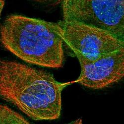
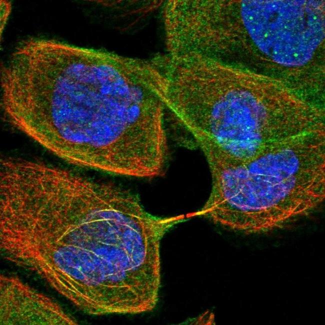
- Immunofluorescent staining of PGM3 in human cell line A-431 shows positivity in cytoplasm, microtubules & nucleus but excluded from the nucleoli. Samples were probed using a PGM3 Polyclonal Antibody (Product # PA5-56249).
Supportive validation
- Submitted by
- Invitrogen Antibodies (provider)
- Main image
- Experimental details
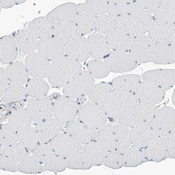
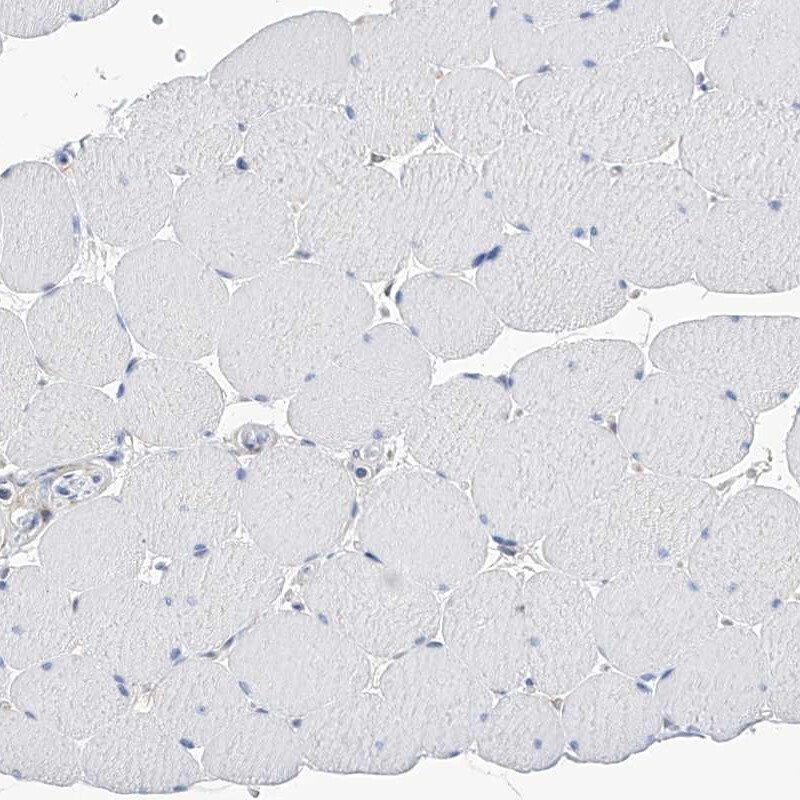
- Immunohistochemical staining of PGM3 in human skeletal muscle using PGM3 Polyclonal Antibody (Product # PA5-56249) shows low expression as expected.
- Submitted by
- Invitrogen Antibodies (provider)
- Main image
- Experimental details
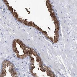
- Immunohistochemical staining of PGM3 in human prostate using PGM3 Polyclonal Antibody (Product # PA5-56249) shows high expression.
Supportive validation
- Submitted by
- Invitrogen Antibodies (provider)
- Main image
- Experimental details
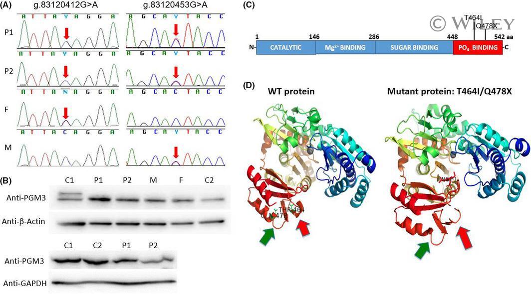
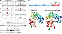
- 2 FIGURE Compound heterozygous PGM3 variants in siblings with IgE-related dermatitis and T-cell lymphopenia. A, Electropherograms showing the Sanger sequencing results of the variant site in PGM3 from gDNA extracted from leukocytes of patients and parents. The variants g. 83120412 G > A and g. 83120453 G > A, compound heterozygous in patients and heterozygous in parents are indicated with red arrow. P1 and P2 are patients, F is the father, and M is the mother. B, Immunoblot analysis of PGM3 in PBMCs (upper panels) and SV40 fibroblast (lower panels) P1, P2 controls (C1 and C2), and the patients' father (F) and mother (M). beta-Actin and GAPDH were used as loading controls. The panels illustrate results from a single experiment, representative of three. C, Representation of PGM3 protein, isoform 1, indicating different domains and the position of the variants T464I and Q478X. D, Protein modeling of WT and mutated PGM3 indicated by green and red arrows' mutations T464I and Q478X [Colour figure can be viewed at wileyonlinelibrary.com ]