Explore
 Validate
 Learn
Learn Western blot
Antibody data
- Antibody Data
- Antigen structure
- References [1]
- Comments [0]
- Validations
- Western blot [4]
- Immunocytochemistry [2]
- Immunoprecipitation [1]
- Immunohistochemistry [6]
- Chromatin Immunoprecipitation [1]
Submit
Validation data
Reference
Comment
Report error
- Product number
- GTX114777 - Provider product page

- Provider
- GeneTex
- Proper citation
- GeneTex Cat#GTX114777, RRID:AB_11164873
- Product name
- SMARCC1 antibody [C2C3], C-term
- Antibody type
- Polyclonal
- Reactivity
- Human, Mouse, Rat
- Host
- Rabbit
Submitted references NKX6.1 functions as a metastatic suppressor through epigenetic regulation of the epithelial-mesenchymal transition.
Li HJ, Yu PN, Huang KY, Su HY, Hsiao TH, Chang CP, Yu MH, Lin YW
Oncogene 2016 Apr 28;35(17):2266-78
Oncogene 2016 Apr 28;35(17):2266-78
No comments: Submit comment
Supportive validation
- Submitted by
- GeneTex (provider)
- Main image
- Experimental details
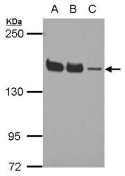
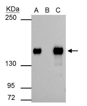
- Sample (30 ?g of whole cell lysate) A: NT2D1 B: IMR32 C: U87-MG 5% SDS PAGE GTX114777 diluted at 1:5000 The HRP-conjugated anti-rabbit IgG antibody (GTX213110-01) was used to detect the primary antibody.

- Submitted by
- GeneTex (provider)
- Main image
- Experimental details
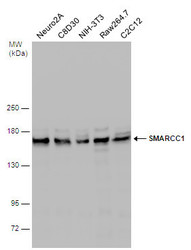
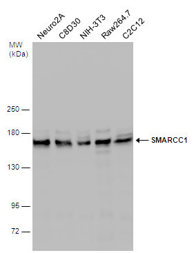
- Various whole cell extracts (30 ?g) were separated by 5% SDS-PAGE, and the membrane was blotted with SMARCC1 antibody [C2C3], C-term (GTX114777) diluted at 1:1000. The HRP-conjugated anti-rabbit IgG antibody (GTX213110-01) was used to detect the primary antibody.
- Submitted by
- GeneTex (provider)
- Main image
- Experimental details
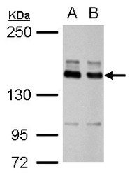
- SMARCC1 antibody detects SMARCC1 protein by Western blot analysis.A. 30 £gg HeLa whole cell lysate/extractB. 30 £gg HepG2 whole cell lysate/extract5 % SDS-PAGESMARCC1 antibody (GTX114777) dilution: 1:10000

- Submitted by
- GeneTex (provider)
- Main image
- Experimental details
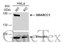
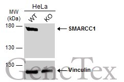
- Wild-type (WT) and SMARCC1 knockout (KO) HeLa cell extracts (30 ?g) were separated by 5% SDS-PAGE, and the membrane was blotted with SMARCC1 antibody [C2C3], C-term (GTX114777) diluted at 1:5000. The HRP-conjugated anti-rabbit IgG antibody (GTX213110-01) was used to detect the primary antibody.
Supportive validation
- Submitted by
- GeneTex (provider)
- Main image
- Experimental details
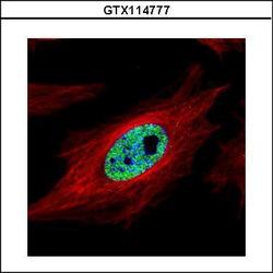
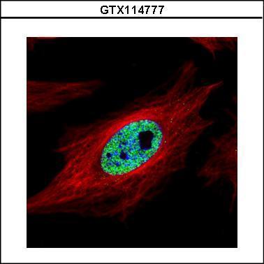
- Confocal immunofluorescence analysis (Olympus FV10i) of paraformaldehyde-fixed HeLa, using SMARCC1(GTX114777) antibody (Green) at 1:500 dilution. Alpha-tubulin filaments were labeled with GTX11304 (Red) at 1:2000.
- Submitted by
- GeneTex (provider)
- Main image
- Experimental details
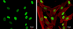
- SMARCC1 antibody [C2C3], C-term detects SMARCC1 protein at nucleus by immunofluorescent analysis.Sample: SK-N-SH cells were fixed in 4% paraformaldehyde at RT for 15 min.Green: SMARCC1 protein stained by SMARCC1 antibody [C2C3], C-term (GTX114777) diluted at 1:500.Red: Phalloidin, a cytoskeleton marker, diluted at 1:200.Scale bar = 10 £gm.

Supportive validation
- Submitted by
- GeneTex (provider)
- Main image

- Experimental details
- SMARCC1 antibody immunoprecipitates SMARCC1 protein in IP experiments. IP Sample: 293T whole cell lysate/extract A : 30 £gg whole cell lysate/extract of SMARCC1 protein expressing 293T cells B : Control with 2.5 £gg of pre-immune rabbit IgG C : Immunoprecipitation of SMARCC1 by 2.5 £gg of SMARCC1 antibody (GTX114777) 5% SDS-PAGE The immunoprecipitated SMARCC1 protein was detected by SMARCC1 antibody (GTX114777) diluted at 1 : 1000. EasyBlot anti-rabbit IgG (HRP) (GTX221666-01) was used as a secondary reagent.
Supportive validation
- Submitted by
- GeneTex (provider)
- Main image
- Experimental details
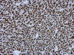
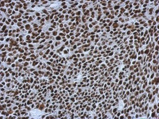
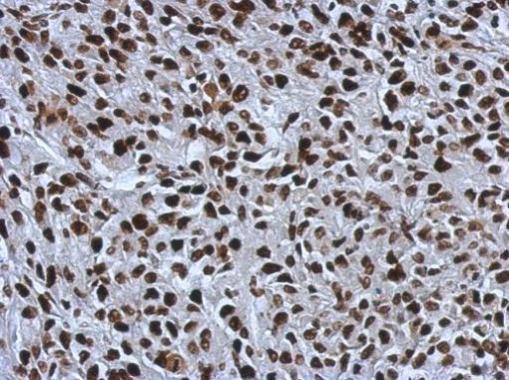
- Immunohistochemical analysis of paraffin-embedded BT483 xenograft, using SMARCC1(GTX114777) antibody at 1:500 dilution.
- Submitted by
- GeneTex (provider)
- Main image
- Experimental details
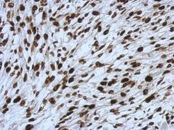
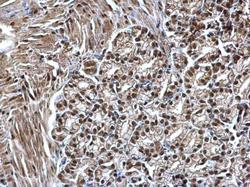
- Immunohistochemical analysis of paraffin-embedded C2C12 xenograft, using SMARCC1(GTX114777) antibody at 1:500 dilution.
- Submitted by
- GeneTex (provider)
- Main image
- Experimental details
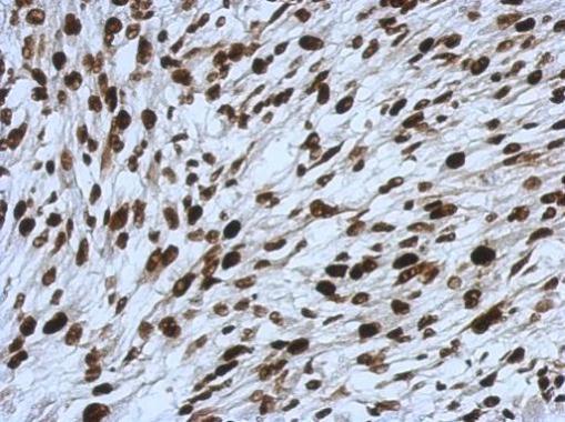
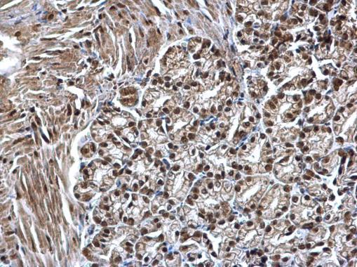
- Immunohistochemical analysis of paraffin-embedded RT2 xenograft, using SMARCC1(GTX114777) antibody at 1:500 dilution.
- Submitted by
- GeneTex (provider)
- Main image
- Experimental details
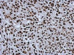
- SMARCC1 antibody [C2C3], C-term detects SMARCC1 protein at nucleus on mouse duodenum by immunohistochemical analysis. Sample: Paraffin-embedded mouse duodenum. SMARCC1 antibody [C2C3], C-term (GTX114777) dilution: 1:500.
- Submitted by
- GeneTex (provider)
- Main image

- Experimental details
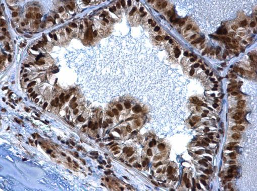
- SMARCC1 antibody [C2C3], C-term detects SMARCC1 protein at nucleus on mouse prostate by immunohistochemical analysis. Sample: Paraffin-embedded mouse prostate. SMARCC1 antibody [C2C3], C-term (GTX114777) dilution: 1:500.
- Submitted by
- GeneTex (provider)
- Main image
- Experimental details
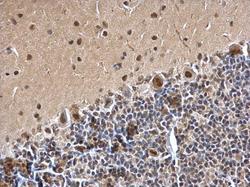
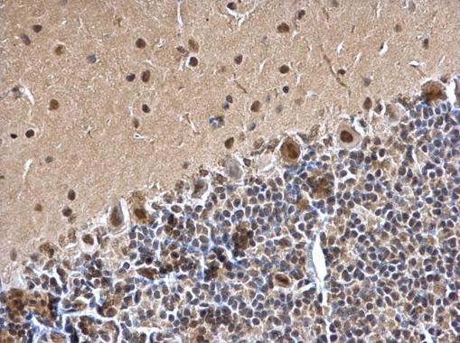
- SMARCC1 antibody [C2C3], C-term detects SMARCC1 protein at nucleus on rat hind brain by immunohistochemical analysis. Sample: Paraffin-embedded rat hind brain. SMARCC1 antibody [C2C3], C-term (GTX114777) dilution: 1:500.
Supportive validation
- Submitted by
- GeneTex (provider)
- Main image
- Experimental details
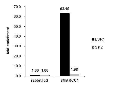
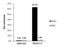
- Cross-linked ChIP was performed with MF-7 chromatin extract treated with B-estradiol (10 nM for 45 min) and 5 £gg of either control rabbit IgG or anti-SMARCC1 antibody. The precipitated DNA was detected by PCR with primer set targeting to ESR1 or Sat2.