 Explore
 Validate
 Learn
Learn Western blot
Antibody data
- Antibody Data
- Antigen structure
- References [0]
- Comments [0]
- Validations
- Western blot [5]
- Immunocytochemistry [7]
- Immunoprecipitation [1]
- Immunohistochemistry [8]
- Chromatin Immunoprecipitation [4]
- Other assay [1]
Submit
Validation data
Reference
Comment
Report error
- Product number
- PA5-30174 - Provider product page

- Provider
- Invitrogen Antibodies
- Product name
- SMARCC1 Polyclonal Antibody
- Antibody type
- Polyclonal
- Antigen
- Recombinant full-length protein
- Description
- Recommended positive controls: HeLa, HepG2, Neuro 2A, C8D30, NIH-3T3, Raw264.7, C2C12. Predicted reactivity: Mouse (96%), Rat (97%). Store product as a concentrated solution. Centrifuge briefly prior to opening the vial.
- Reactivity
- Human, Mouse, Rat
- Host
- Rabbit
- Isotype
- IgG
- Vial size
- 100 μL
- Concentration
- 0.77 mg/mL
- Storage
- Store at 4°C short term. For long term storage, store at -20°C, avoiding freeze/thaw cycles.
No comments: Submit comment
Supportive validation
- Submitted by
- Invitrogen Antibodies (provider)
- Main image
- Experimental details
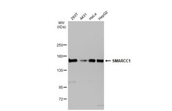
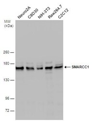
- Western Blot using SMARCC1 Polyclonal Antibody (Product # PA5-30174). Various whole cell extracts (30 µg) were separated by 5% SDS-PAGE, and the membrane was blotted with SMARCC1 Polyclonal Antibody (Product # PA5-30174) diluted at 1:2,000. The HRP-conjugated anti-rabbit IgG antibody was used to detect the primary antibody.
- Submitted by
- Invitrogen Antibodies (provider)
- Main image
- Experimental details
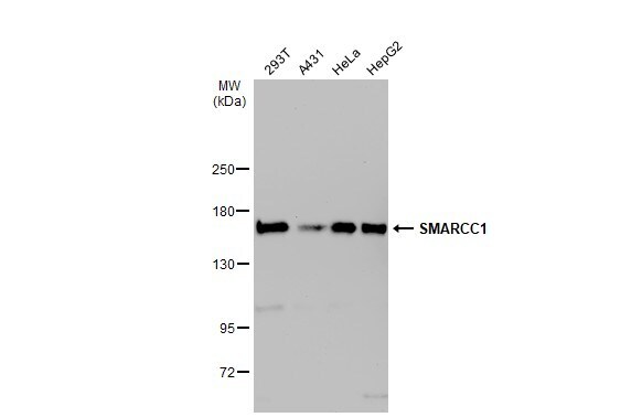
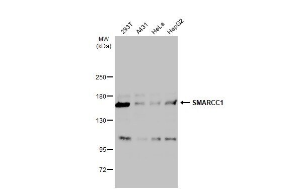
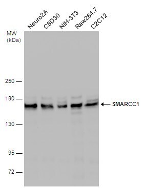
- Western Blot analysis of SMARCC1 was performed by separating 30 µg of various whole cell extracts by 5% SDS-PAGE. Proteins were transferred to a membrane and probed with a SMARCC1 Polyclonal Antibody (Product # PA5-30174) at a dilution of 1:2000 and a HRP-conjugated anti-rabbit IgG secondary antibody.
- Submitted by
- Invitrogen Antibodies (provider)
- Main image
- Experimental details
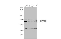
- Western Blot analysis of SMARCC1 was performed by separating 30 µg of various whole cell extracts by 5% SDS-PAGE. Proteins were transferred to a membrane and probed with a SMARCC1 Polyclonal Antibody (Product # PA5-30174) at a dilution of 1:1000 and a HRP-conjugated anti-rabbit IgG secondary antibody.
- Submitted by
- Invitrogen Antibodies (provider)
- Main image
- Experimental details
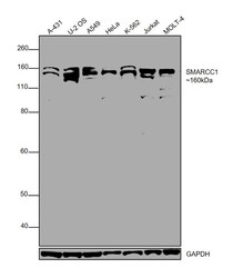
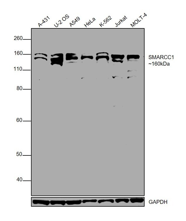
- Western blot was performed using Anti-SMARCC1 Polyclonal Antibody (Product # PA5-30174) and a 160 kDa band corresponding to SMARCC1 was observed in all the tested cell models. Modified whole cell lysate (1% SDS) (30 µg lysate) of A-431 (Lane 1), U-2 OS (Lane 2), A549 (Lane 3), HeLa (Lane 4), K-562 (Lane 5), Jurkat (Lane 6) and MOLT-4 (Lane 7) were electrophoresed using NuPAGE® 10 % Bis-Tris gel (Product # NP0302BOX). Resolved proteins were then transferred onto a nitrocellulose membrane (Product # IB23001) by wet transfer. The blot was probed with the primary antibody (1:1,000 dilution) and detected by chemiluminescence with Goat anti-Rabbit IgG (Heavy Chain), Superclonal™ Recombinant Secondary Antibody, HRP conjugate (Product # A27036, 1:4,000 dilution) using the iBright FL 1000 (Product # A32752). Chemiluminescent detection was performed using SuperSignal™ West Dura Extended Duration Substrate (Product # 34076).
- Submitted by
- Invitrogen Antibodies (provider)
- Main image

- Experimental details
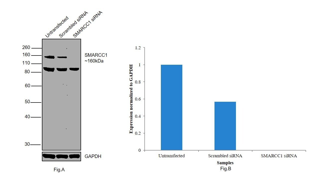
- Knockdown of SMARCC1 was achieved by transfecting HeLa with SMARCC1 specific siRNAs (Silencer® select Products # s13145, s13146). Western blot analysis (Fig. a) was performed using whole cell extracts from the SMARCC1 knockdown cells (Lane 3), non-specific scrambled siRNA transfected cells (Lane 2) and untransfected cells (Lane 1). The blot was probed with SMARCC1 Polyclonal Antibody (Product # PA5-30174, 1:1,000 dilution) and Goat anti-Rabbit IgG (Heavy Chain) Superclonal™ Secondary Antibody, HRP conjugate (Product # A27036, 0.25 µg/mL, 1:4,000 dilution). Densitometric analysis of this western blot is shown in histogram (Fig. b). Loss of signal upon siRNA mediated knock down confirms that antibody is specific to SMARCC1. (Note: Few uncharacterized bands were observed at ~ 90 kDa).
Supportive validation
- Submitted by
- Invitrogen Antibodies (provider)
- Main image
- Experimental details
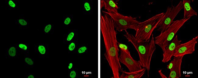
- Immunofluorescent analysis of SMARCC1 in paraformaldehyde-fixed HeLa cells using a SMARCC1 polyclonal antibody (Product # PA5-30174) (Green) at a 1:500 dilution. Alpha-tubulin filaments were labeled with Product # PA5-29281 (Red) at a 1:2000.

- Submitted by
- Invitrogen Antibodies (provider)
- Main image

- Experimental details
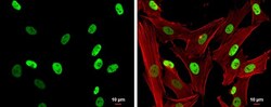
- Immunocytochemistry-Immunofluorescence analysis of SMARCC1 was performed in SK-N-SH cells fixed in 4% paraformaldehyde at RT for 15 min. Green: SMARCC1 Polyclonal Antibody (Product # PA5-30174) diluted at 1:500. Red: Phalloidin, a cytoskeleton marker. Scale bar = 10 µm.

- Submitted by
- Invitrogen Antibodies (provider)
- Main image

- Experimental details
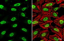
- SMARCC1 Polyclonal Antibody detects SMARCC1 protein at nucleus by immunofluorescent analysis. Sample: HeLa cells were fixed in 4% paraformaldehyde at RT for 15 min. Green: SMARCC1 stained by SMARCC1 Polyclonal Antibody (Product # PA5-30174) diluted at 1:500. Red: phalloidin, a cytoskeleton marker, diluted at 1:200. Scale bar= 10 µm.

- Submitted by
- Invitrogen Antibodies (provider)
- Main image
- Experimental details
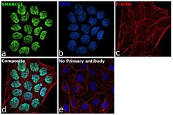
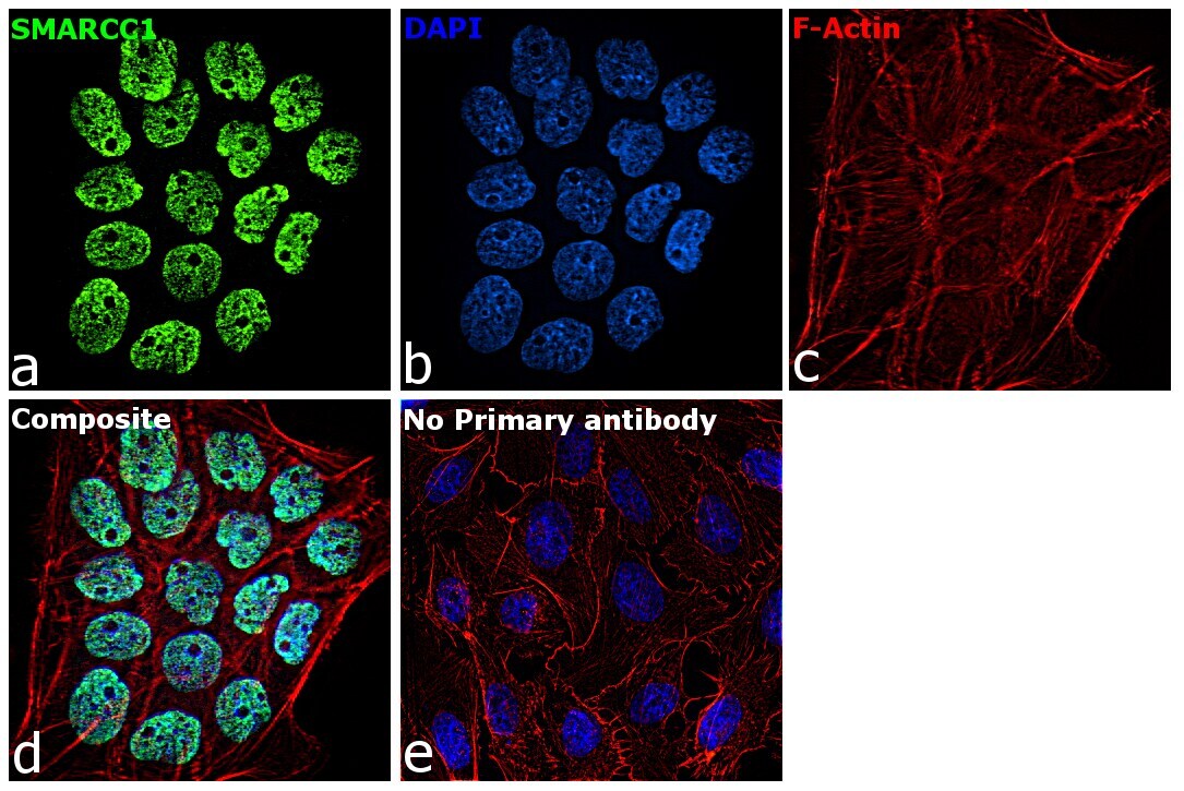
- Immunofluorescence analysis of SMARCC1 was performed using 70% confluent log phase A-431 cells. The cells were fixed with 4% paraformaldehyde for 10 minutes, permeabilized with 0.1% Triton™ X-100 for 15 minutes, and blocked with 2% BSA for 1 hour at room temperature. The cells were labeled with SMARCC1 Polyclonal Antibody (Product # PA5-30174) at 5 µg/mL in 0.1% BSA, incubated at 4 degree Celsius overnight and then labeled with Goat anti-Rabbit IgG (H+L) Superclonal™ Recombinant Secondary Antibody, Alexa Fluor® 488 conjugate (Product # A27034) at a dilution of 1:2000 for 45 minutes at room temperature (Panel a: green). Nuclei (Panel b: blue) were stained with ProLong™ Diamond Antifade Mountant with DAPI (Product # P36962). F-actin (Panel c: red) was stained with Rhodamine Phalloidin (Product # R415, 1:300). Panel d represents the merged image showing nuclear localization. Panel e represents control cells with no primary antibody to assess background. The images were captured at 60X magnification.
- Submitted by
- Invitrogen Antibodies (provider)
- Main image
- Experimental details
- SMARCC1 Polyclonal Antibody detects SMARCC1 protein at nucleus by immunofluorescent analysis. Sample: HeLa cells were fixed in 4% paraformaldehyde at RT for 15 min. Green: SMARCC1 stained by SMARCC1 Polyclonal Antibody (Product # PA5-30174) diluted at 1:500. Red: phalloidin, a cytoskeleton marker, diluted at 1:200. Scale bar= 10 µm.

- Submitted by
- Invitrogen Antibodies (provider)
- Main image

- Experimental details
- Immunofluorescence analysis of SMARCC1 was performed using 70% confluent log phase A-431 cells. The cells were fixed with 4% paraformaldehyde for 10 minutes, permeabilized with 0.1% Triton™ X-100 for 15 minutes, and blocked with 2% BSA for 1 hour at room temperature. The cells were labeled with SMARCC1 Polyclonal Antibody (Product # PA5-30174) at 5 µg/mL in 0.1% BSA, incubated at 4 degree Celsius overnight and then labeled with Goat anti-Rabbit IgG (Heavy Chain) Superclonal™ Recombinant Secondary Antibody, Alexa Fluor® 488 conjugate (Product # A27034) at a dilution of 1:2000 for 45 minutes at room temperature (Panel a: green). Nuclei (Panel b: blue) were stained with ProLong™ Diamond Antifade Mountant with DAPI (Product # P36962). F-actin (Panel c: red) was stained with Rhodamine Phalloidin (Product # R415, 1:300). Panel d represents the merged image showing nuclear localization. Panel e represents control cells with no primary antibody to assess background. The images were captured at 60X magnification.

- Submitted by
- Invitrogen Antibodies (provider)
- Main image
- Experimental details
- Immunocytochemistry-Immunofluorescence analysis of SMARCC1 was performed in SK-N-SH cells fixed in 4% paraformaldehyde at RT for 15 min. Green: SMARCC1 Polyclonal Antibody (Product # PA5-30174) diluted at 1:500. Red: Phalloidin, a cytoskeleton marker. Scale bar = 10 µm.
Supportive validation
- Submitted by
- Invitrogen Antibodies (provider)
- Main image
- Experimental details
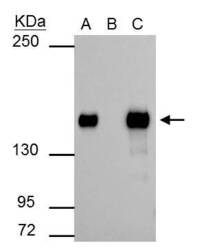
- SMARCC1 antibody immunoprecipitates SMARCC1 protein in IP experiments. IP Sample: 293T whole cell lysate/extract A : 30 µg whole cell lysate/extract of SMARCC1 protein expressing 293T cells B : Control with 2.5 µg of pre-immune rabbit IgG C : Immunoprecipitation of SMARCC1 by 2.5 µg of SMARCC1 antibody (Product # PA5-30174) 5% SDS-PAGE The immunoprecipitated SMARCC1 protein was detected by SMARCC1 antibody (Product # PA5-30174) diluted at 1:1,000. Anti-rabbit IgG (HRP) was used as a secondary reagent.

Supportive validation
- Submitted by
- Invitrogen Antibodies (provider)
- Main image
- Experimental details
- SMARCC1 Polyclonal Antibody detects SMARCC1 protein at nucleus on mouse duodenum by immunohistochemical analysis. Sample: Paraffin-embedded mouse duodenum. SMARCC1 Polyclonal Antibody (Product # PA5-30174) dilution: 1:500. Antigen Retrieval: EDTA based buffer, pH 8.0, 15 min.

- Submitted by
- Invitrogen Antibodies (provider)
- Main image

- Experimental details
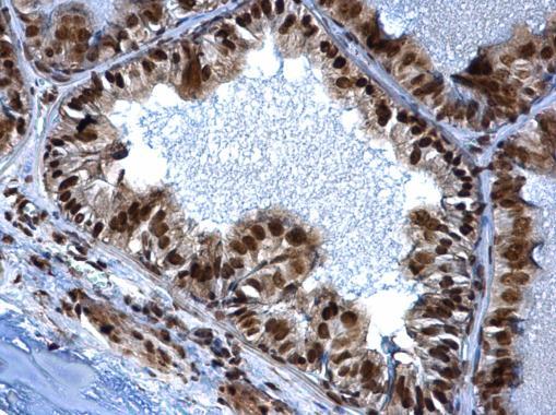
- SMARCC1 Polyclonal Antibody detects SMARCC1 protein at nucleus on mouse prostate by immunohistochemical analysis. Sample: Paraffin-embedded mouse prostate. SMARCC1 Polyclonal Antibody (Product # PA5-30174) dilution: 1:500. Antigen Retrieval: EDTA based buffer, pH 8.0, 15 min.
- Submitted by
- Invitrogen Antibodies (provider)
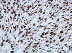
- Main image
- Experimental details
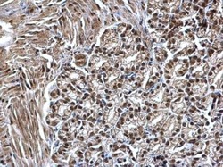
- Immunohistochemical analysis of paraffin-embedded RT2 xenograft, using SMARCC1 (Product # PA5-30174) antibody at 1:500 dilution. Antigen Retrieval: EDTA based buffer, pH 8.0, 15 min.

- Submitted by
- Invitrogen Antibodies (provider)
- Main image
- Experimental details
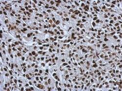
- Immunohistochemical analysis of paraffin-embedded BT483 xenograft, using SMARCC1 (Product # PA5-30174) antibody at 1:500 dilution. Antigen Retrieval: EDTA based buffer, pH 8.0, 15 min.

- Submitted by
- Invitrogen Antibodies (provider)
- Main image
- Experimental details
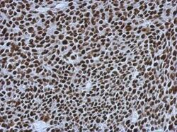
- Immunohistochemical analysis of paraffin-embedded C2C12 xenograft, using SMARCC1 (Product # PA5-30174) antibody at 1:500 dilution. Antigen Retrieval: EDTA based buffer, pH 8.0, 15 min.

- Submitted by
- Invitrogen Antibodies (provider)
- Main image
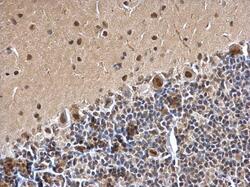
- Experimental details
- SMARCC1 Polyclonal Antibody detects SMARCC1 protein at nucleus on rat hind brain by immunohistochemical analysis. Sample: Paraffin-embedded rat hind brain. SMARCC1 Polyclonal Antibody (Product # PA5-30174) dilution: 1:500. Antigen Retrieval: EDTA based buffer, pH 8.0, 15 min.
- Submitted by
- Invitrogen Antibodies (provider)
- Main image

- Experimental details
- SMARCC1 Polyclonal Antibody detects SMARCC1 protein at nucleus on mouse prostate by immunohistochemical analysis. Sample: Paraffin-embedded mouse prostate. SMARCC1 Polyclonal Antibody (Product # PA5-30174) dilution: 1:500. Antigen Retrieval: EDTA based buffer, pH 8.0, 15 min.

- Submitted by
- Invitrogen Antibodies (provider)
- Main image

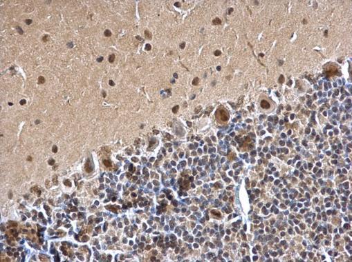
- Experimental details
- SMARCC1 Polyclonal Antibody detects SMARCC1 protein at nucleus on rat hind brain by immunohistochemical analysis. Sample: Paraffin-embedded rat hind brain. SMARCC1 Polyclonal Antibody (Product # PA5-30174) dilution: 1:500. Antigen Retrieval: EDTA based buffer, pH 8.0, 15 min.

Supportive validation
- Submitted by
- Invitrogen Antibodies (provider)
- Main image
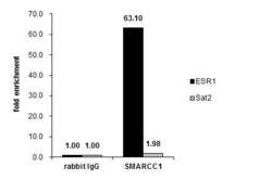
- Experimental details
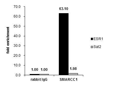
- Cross-linked ChIP was performed with MF-7 chromatin extract treated with B-estradiol (10 nM for 45 min) and 5 µg of either control rabbit IgG or SMARCC1 Polyclonal Antibody (Product # PA5-30174). The precipitated DNA was detected by PCR with primer set targeting to ESR1 or Sat2.

- Submitted by
- Invitrogen Antibodies (provider)
- Main image
- Experimental details
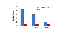
- Chromatin Immunoprecipitation (ChIP) assay of endogenous SMARCC1 protein using Anti-SMARCC1 Antibody: ChIP was performed using Anti-SMARCC1 Rabbit Polyclonal Antibody (Product # PA5-30174, 5 µg) on sheared chromatin from HeLa cells using the MAGnify ChIP System kit (Product # 49-2024). Normal Rabbit IgG was used as a negative IP control. The purified DNA was analyzed by qPCR using primers binding to pS2 and CCND1 promoters and SAT2 satellite repeats. Data is presented as fold enrichment of the antibody signal versus the negative control IgG using the comparative CT method.

- Submitted by
- Invitrogen Antibodies (provider)
- Main image

- Experimental details
- Cross-linked ChIP was performed with MF-7 chromatin extract treated with B-estradiol (10 nM for 45 min) and 5 µg of either control rabbit IgG or SMARCC1 Polyclonal Antibody (Product # PA5-30174). The precipitated DNA was detected by PCR with primer set targeting to ESR1 or Sat2.
Supportive validation
- Submitted by
- Invitrogen Antibodies (provider)
- Main image

- Experimental details
- SMARCC1 antibody immunoprecipitates SMARCC1 protein in IP experiments. IP Sample: 293T whole cell lysate/extract A : 30 µg whole cell lysate/extract of SMARCC1 protein expressing 293T cells B : Control with 2.5 µg of pre-immune rabbit IgG C : Immunoprecipitation of SMARCC1 by 2.5 µg of SMARCC1 antibody (Product # PA5-30174) 5% SDS-PAGE The immunoprecipitated SMARCC1 protein was detected by SMARCC1 antibody (Product # PA5-30174) diluted at 1:1,000. Anti-rabbit IgG (HRP) was used as a secondary reagent.
