 Explore
 Validate
 Learn
Learn Western blot
 ELISA
ELISAAntibody data
- Antibody Data
- Antigen structure
- References [3]
- Comments [0]
- Validations
- Western blot [1]
- Immunohistochemistry [1]
Submit
Validation data
Reference
Comment
Report error
- Product number
- NB100-57094 - Provider product page

- Provider
- Novus Biologicals
- Proper citation
- Novus Cat#NB100-57094, RRID:AB_2199372
- Product name
- Goat Polyclonal TBC1D4 Antibody
- Antibody type
- Polyclonal
- Description
- Immunogen affinity purified.
- Reactivity
- Human
- Host
- Goat
- Antigen sequence
DDPEKIEERKKSK- Isotype
- IgG
- Vial size
- 0.1 mg
- Concentration
- 0.5 mg/ml
- Storage
- Store at -20C. Avoid freeze-thaw cycles.
Submitted references Regulation of glucose transporter 4 translocation by the Rab guanosine triphosphatase-activating protein AS160/TBC1D4: role of phosphorylation and membrane association.
Insulin-stimulated phosphorylation of the Akt substrate AS160 is impaired in skeletal muscle of type 2 diabetic subjects.
Insulin-stimulated phosphorylation of the Akt substrate AS160 is impaired in skeletal muscle of type 2 diabetic subjects.
Stöckli J, Davey JR, Hohnen-Behrens C, Xu A, James DE, Ramm G
Molecular endocrinology (Baltimore, Md.) 2008 Dec;22(12):2703-15
Molecular endocrinology (Baltimore, Md.) 2008 Dec;22(12):2703-15
Insulin-stimulated phosphorylation of the Akt substrate AS160 is impaired in skeletal muscle of type 2 diabetic subjects.
Karlsson HK, Zierath JR, Kane S, Krook A, Lienhard GE, Wallberg-Henriksson H
Diabetes 2005 Jun;54(6):1692-7
Diabetes 2005 Jun;54(6):1692-7
Insulin-stimulated phosphorylation of the Akt substrate AS160 is impaired in skeletal muscle of type 2 diabetic subjects.
Karlsson HK, Zierath JR, Kane S, Krook A, Lienhard GE, Wallberg-Henriksson H
Diabetes 2005 Jun;54(6):1692-7
Diabetes 2005 Jun;54(6):1692-7
No comments: Submit comment
Supportive validation
- Submitted by
- Novus Biologicals (provider)
- Main image
- Experimental details
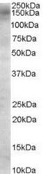
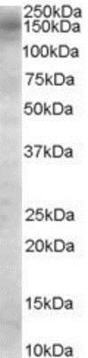
- Western Blot: TBC1D4 Antibody [NB100-57094] - (0.1ug/ml) staining of Daudi cell lysate (35ug protein in RIPA buffer). Primary incubation was 1 hour. Detected by chemiluminescence.
Supportive validation
- Submitted by
- Novus Biologicals (provider)
- Main image
- Experimental details
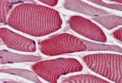
- Immunohistochemistry-Paraffin: TBC1D4 Antibody [NB100-57094] - (3.8ug/ml) staining of Human Skeletal Muscle. Steamed antigen retrieval with citrate buffer pH 6, AP-staining.
