Explore
 Validate
 Learn
Learn Western blot
Antibody data
- Antibody Data
- Antigen structure
- References [0]
- Comments [0]
- Validations
- Western blot [4]
- Immunocytochemistry [1]
- Immunohistochemistry [1]
Submit
Validation data
Reference
Comment
Report error
- Product number
- RQ5339 - Provider product page

- Provider
- NSJ Bioreagents
- Product name
- IRF1 Antibody / Interferon regulatory factor 1
- Antibody type
- Monoclonal
- Description
- This highly specific IRF1 antibody is suitable for use in Immunohistochemistry/Western blot applications with human and rat samples.
- Reactivity
- Human, Rat
- Host
- Rabbit
- Conjugate
- Unconjugated
- Antibody clone number
- DHE-9
- Vial size
- 100 ul
- Concentration
- Antibody in PBS with 0.02% sodium azide, 50% glycerol and 0.4-0.5mg/ml BSA
- Storage
- Store the IRF1 antibody at -20oC.
No comments: Submit comment
Supportive validation
- Submitted by
- NSJ Bioreagents (provider)
- Main image
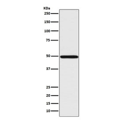
- Experimental details
- Western blot testing of human Jurkat cell lysate with IRF1 antibody. Expected molecular weight: ~37 kDa (unmodified), 45-50 kDa (modified).
- Submitted by
- NSJ Bioreagents (provider)
- Main image
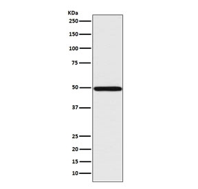
- Experimental details
- Western blot testing of human Jurkat cell lysate with IRF1 antibody. Expected molecular weight: ~37 kDa (unmodified), 45-50 kDa (modified).

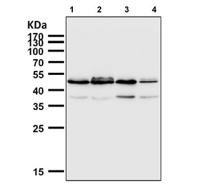
- Submitted by
- NSJ Bioreagents (provider)
- Main image
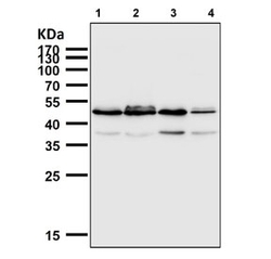
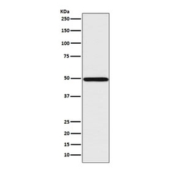
- Experimental details
- Western blot testing of human 1) HaCaT, 2) Raji, 3) K562 and 4) Daudi cell lysate with IRF1 antibody. Expected molecular weight: ~37 kDa (unmodified), 45-50 kDa (modified).
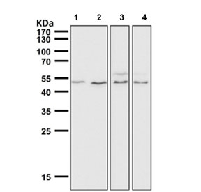
- Submitted by
- NSJ Bioreagents (provider)
- Main image
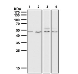
- Experimental details
- Western blot testing of 1) mouse heart, 2) mouse liver, 3) mouse skin and 4) rat skin tissue lysate with IRF1 antibody. Expected molecular weight: ~37 kDa (unmodified), 45-50 kDa (modified).
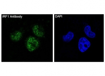
Supportive validation
- Submitted by
- NSJ Bioreagents (provider)
- Main image
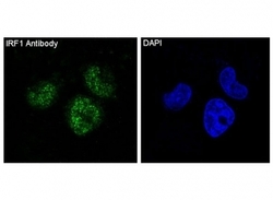
- Experimental details
- Immunofluorescent staining of FFPE human HeLa cells with IRF1 antibody (green) and DAPI nuclear stain (blue). HIER: steam section in pH6 citrate buffer for 20 min.
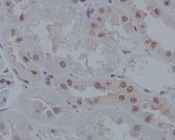
Supportive validation
- Submitted by
- NSJ Bioreagents (provider)
- Main image

- Experimental details
- IHC staining of FFPE rat kidney tissue with IRF1 antibody. HIER: boil tissue sections in pH6, 10mM citrate buffer, for 10-20 min and allow to cool before testing.