Explore
 Validate
 Learn
Learn Western blot
 ELISA
ELISAAntibody data
- Antibody Data
- Antigen structure
- References [2]
- Comments [0]
- Validations
- Western blot [1]
Submit
Validation data
Reference
Comment
Report error
- Product number
- PB10063 - Provider product page

- Provider
- Boster Biological Technology
- Product name
- Anti-FGF21 Antibody Picoband™
- Antibody type
- Polyclonal
- Description
- Polyclonal antibody for FGF-21/FGF21 detection. Host: Rabbit.Size: 100μg/vial. Tested applications: ELISA. Reactive species: Human. FGF-21/FGF21 information: Molecular Weight: 22300 MW; Subcellular Localization: Secreted .
- Reactivity
- Human, Mouse, Rat
- Host
- Rabbit
- Vial size
- 100μg/vial
- Concentration
- Add 0.2ml of distilled water will yield a concentration of 500ug/ml.
- Storage
- At -20°C for one year. After reconstitution, at 4°C for one month. It can also be aliquoted and stored frozen at -20°C for a longer time. Avoid repeated freezing and thawing.
- Handling
- Add 0.2ml of distilled water will yield a concentration of 500ug/ml.
Submitted references Ethyl acetate extract of sappanwood alleviates experimental atherosclerosis in rats through changes in FGF21 and SREBP-2 expression.
Expression of Fibroblast Growth Factor 21 and β-Klotho Regulates Hepatic Fibrosis through the Nuclear Factor-κB and c-Jun N-Terminal Kinase Pathways.
Li Q, Wang H, Zhang C, Tong R, Chen H, Qie R
International journal of clinical and experimental pathology 2020;13(2):220-229
International journal of clinical and experimental pathology 2020;13(2):220-229
Expression of Fibroblast Growth Factor 21 and β-Klotho Regulates Hepatic Fibrosis through the Nuclear Factor-κB and c-Jun N-Terminal Kinase Pathways.
Lee KJ, Jang YO, Cha SK, Kim MY, Park KS, Eom YW, Baik SK
Gut and liver 2018 Jul 15;12(4):449-456
Gut and liver 2018 Jul 15;12(4):449-456
No comments: Submit comment
Supportive validation
- Submitted by
- Boster Biological Technology (provider)
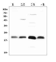
- Main image
- Experimental details
- Western blot analysis of FGF21 using anti-FGF21 antibody (PB10063). Electrophoresis was performed on a 5-20% SDS-PAGE gel at 70V (Stacking gel) / 90V (Resolving gel) for 2-3 hours. The sample well of each lane was loaded with 50ug of sample under reducing conditions. Lane 1: human placenta tissue lysates, After Electrophoresis, proteins were transferred to a Nitrocellulose membrane at 150mA for 50-90 minutes. Blocked the membrane with 5% Non-fat Milk/ TBS for 1.5 hour at RT. The membrane was incubated with rabbit anti-FGF21 antigen affinity purified polyclonal antibody (Catalog # PB10063) at 0.5 μg/mL overnight at 4°C, then washed with TBS-0.1%Tween 3 times with 5 minutes each and probed with a goat anti-rabbit IgG-HRP secondary antibody at a dilution of 1:10000 for 1.5 hour at RT. The signal is developed using an Enhanced Chemiluminescent detection (ECL) kit (Catalog # EK1002) with Tanon 5200 system. A specific band was detected for FGF21 at approximately 22KD. The expected band size for FGF21 is at 22KD.
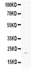
- Additional image