 Explore
 Validate
 Learn
Learn Western blot
 Immunocytochemistry
Antibody data
- Antibody Data
- Antigen structure
- References [1]
- Comments [0]
- Validations
- Immunocytochemistry [3]
- Other assay [1]
Submit
Validation data
Reference
Comment
Report error
- Product number
- MA5-14804 - Provider product page

- Provider
- Invitrogen Antibodies
- Product name
- SETD8 Monoclonal Antibody (B.540.4)
- Antibody type
- Monoclonal
- Antigen
- Synthetic peptide
- Description
- It is not recommended to aliquot this antibody.
- Reactivity
- Human, Mouse, Rat
- Host
- Rabbit
- Isotype
- IgG
- Antibody clone number
- B.540.4
- Vial size
- 100 μL
- Concentration
- 25 μg/mL
- Storage
- -20°C
Submitted references USP29 Deubiquitinates SETD8 and Regulates DNA Damage-Induced H4K20 Monomethylation and 53BP1 Focus Formation.
Hernández-Reyes Y, Paz-Cabrera MC, Freire R, Smits VAJ
Cells 2022 Aug 11;11(16)
Cells 2022 Aug 11;11(16)
No comments: Submit comment
Supportive validation
- Submitted by
- Invitrogen Antibodies (provider)
- Main image
- Experimental details
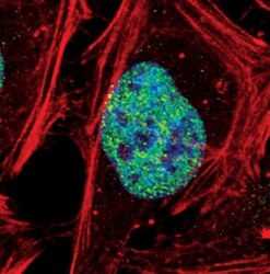
- Immunofluorescent analysis of SETD8 in HeLa cells, treated with 0.5% Triton X-100 (10 minutes prior to fixation), using a SETD8 monoclonal antibody (Product # MA5-14804) (green). Actin filaments are labeled with a fluorescent red phalloidin. DNA is labeled using a fluorescent blue dye.
- Submitted by
- Invitrogen Antibodies (provider)
- Main image

- Experimental details
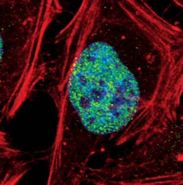
- Immunofluorescent analysis of SETD8 in HeLa cells using a SETD8 monoclonal antibody (Product # MA5-14804) (green). Actin filaments are labeled with a fluorescent red phalloidin. DNA is labeled using a fluorescent blue dye.
- Submitted by
- Invitrogen Antibodies (provider)
- Main image
- Experimental details
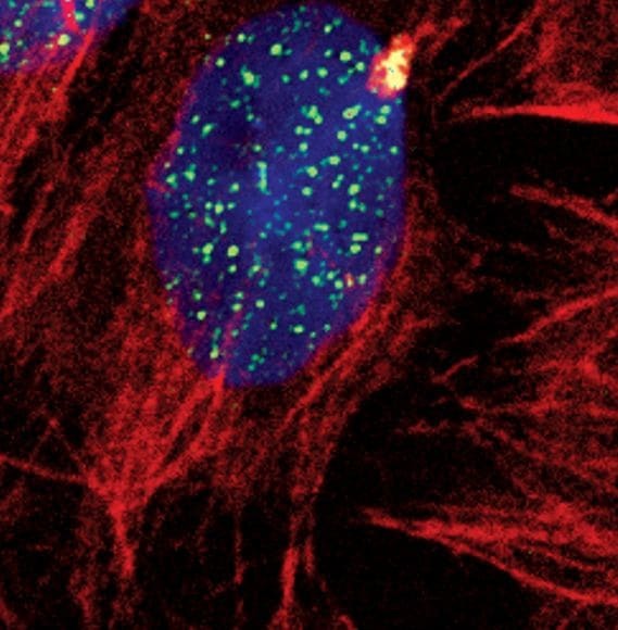
- Immunofluorescent analysis of SETD8 in HeLa cells using a SETD8 monoclonal antibody (Product # MA5-14804) (green). Actin filaments are labeled with a fluorescent red phalloidin. DNA is labeled using a fluorescent blue dye.

Supportive validation
- Submitted by
- Invitrogen Antibodies (provider)
- Main image
- Experimental details
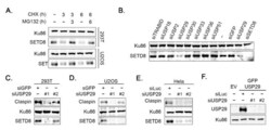
- Depletion of USP29 affects SETD8 protein levels. ( A ) 293T cells and U2OS cells were treated with MG132 and/or CHX for 3 or 6 h. WCEs were analyzed by Western blot using the indicated antibodies. ( B ) Example of screening for ubiquitin hydrolases regulating SETD8 levels. U2OS cells were transfected with the specified siRNA oligonucleotides. Then, 48 h later, cells were lysed, and extracts were analyzed by Western blot with the indicated antibodies. ( C ) The 293T cells were transfected with GFP or the indicated USP29 siRNA oligonucleotides, lysed, and subsequently analyzed by Western blot using the indicated antibodies. ( D ) As in ( C ), but in U2OS cells. ( E ) HeLa cells were transfected with Luciferase (Luc) or the indicated USP29 siRNA oligonucleotides. WCEs were analyzed by Western blot using the indicated antibodies. ( F ) U2OS cells were transfected with an empty vector (EV) or GFP-USP29 and thereafter transfected with Luc or USP29 siRNA oligonucleotides. Extracts were analyzed by Western blot analysis with the indicated antibodies.
