Explore
 Validate
 Learn
Learn Western blot
 ELISA
ELISA Immunocytochemistry
Antibody data
- Antibody Data
- Antigen structure
- References [0]
- Comments [0]
- Validations
- Immunocytochemistry [3]
- Immunohistochemistry [2]
Submit
Validation data
Reference
Comment
Report error
- Product number
- PA5-121097 - Provider product page

- Provider
- Invitrogen Antibodies
- Product name
- BCAT2 Polyclonal Antibody
- Antibody type
- Polyclonal
- Antigen
- Recombinant protein fragment
- Description
- Positive test controls include: MCF7, Jurkat, Mouse brain, Rat kidney. The target is usually found in the following locations: Cytoplasm, Mitochondrion. Immunogen sequence: ASSSFKAADL QLEMTQKPHK KPGPGEPLVF GKTFTDHMLM VEWNDKGWGQ PRIQPFQNLT LHPASSSLHY SLQLFEGMKA FKGKDQQVRL FRPWLNMDRM LRSAMRLCLP SFDKLELLEC IRRLIEVDKD WVPDAAGTSL YVRPVLIGNE PSLGVSQPTR ALLFVILCPV GAYFPGGSVT PVSLLADPAF IRAWVGGVGN YKLGGNYGPT VLVQQEALKR GCEQVLWLYG PDHQLTEVGT MNIFVYWTHE DGVLELVTPP LNGVILPGVV RQSLLDMAQT WGEFRVVERT ITMKQLLRAL EEGRVREVFG SGTACQVCPV HRILYKDRNL HIPTMENGPE LILRFQKELK EIQYGIRAHE WMFPV
- Reactivity
- Human, Mouse, Rat
- Host
- Rabbit
- Isotype
- IgG
- Vial size
- 100 μL
- Concentration
- 1.08 mg/mL
- Storage
- -20°C, Avoid Freeze/Thaw Cycles
No comments: Submit comment
Supportive validation
- Submitted by
- Invitrogen Antibodies (provider)
- Main image
- Experimental details
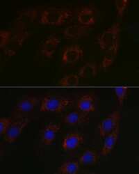
- Immunocytochemical analysis of BCAT2 in NIH-3T3 cells using a BCAT2 Polyclonal antibody (Product # PA5-121097). Blue: DAPI for nuclear staining.

- Submitted by
- Invitrogen Antibodies (provider)
- Main image

- Experimental details
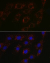
- Immunocytochemical analysis of BCAT2 in NIH-3T3 cells using a BCAT2 Polyclonal antibody (Product # PA5-121097). Blue: DAPI for nuclear staining.
- Submitted by
- Invitrogen Antibodies (provider)
- Main image
- Experimental details
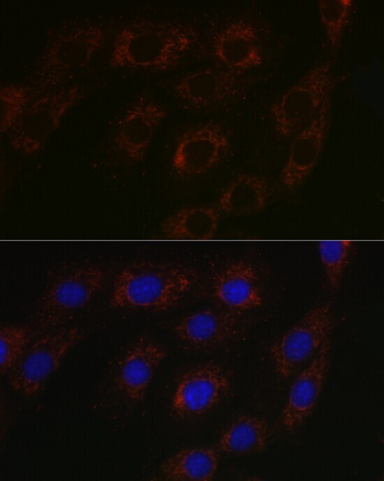
- Immunofluorescence analysis of BCAT2 in NIH-3T3 cells. Samples were incubated with BCAT2 Polyclonal antibody (Product # PA5-121097) using a dilution of 1:100 (40x lens). Blue: DAPI for nuclear staining.

Supportive validation
- Submitted by
- Invitrogen Antibodies (provider)
- Main image
- Experimental details
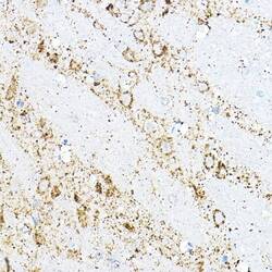
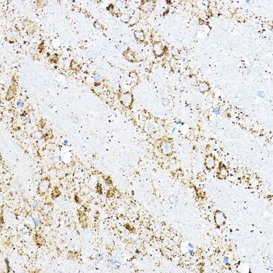
- Immunohistochemistry analysis of BCAT2 in paraffin-embedded rat brain. Samples were incubated with BCAT2 Polyclonal antibody (Product # PA5-121097) using a dilution of 1:100 (40x lens). Perform high pressure antigen retrieval with 10 mM citrate buffer pH 6.0 before commencing with IHC staining protocol.
- Submitted by
- Invitrogen Antibodies (provider)
- Main image
- Experimental details
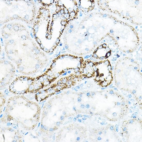
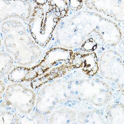
- Immunohistochemistry analysis of BCAT2 in paraffin-embedded mouse kidney. Samples were incubated with BCAT2 Polyclonal antibody (Product # PA5-121097) using a dilution of 1:100 (40x lens). Perform high pressure antigen retrieval with 10 mM citrate buffer pH 6.0 before commencing with IHC staining protocol.