Explore
 Validate
 Learn
Learn Western blot
Antibody data
- Antibody Data
- Antigen structure
- References [1]
- Comments [0]
- Validations
- Western blot [3]
- Immunocytochemistry [2]
- Immunohistochemistry [2]
- Other assay [1]
Submit
Validation data
Reference
Comment
Report error
- Product number
- PA5-28992 - Provider product page

- Provider
- Invitrogen Antibodies
- Product name
- eIF2b alpha Polyclonal Antibody
- Antibody type
- Polyclonal
- Antigen
- Recombinant full-length protein
- Description
- Recommended positive controls: 293T, A431, Jurkat, Raji. Predicted reactivity: Mouse (94%), Rat (92%), Bovine (95%). Store product as a concentrated solution. Centrifuge briefly prior to opening the vial.
- Reactivity
- Human, Mouse
- Host
- Rabbit
- Isotype
- IgG
- Vial size
- 100 μL
- Concentration
- 1 mg/mL
- Storage
- Store at 4°C short term. For long term storage, store at -20°C, avoiding freeze/thaw cycles.
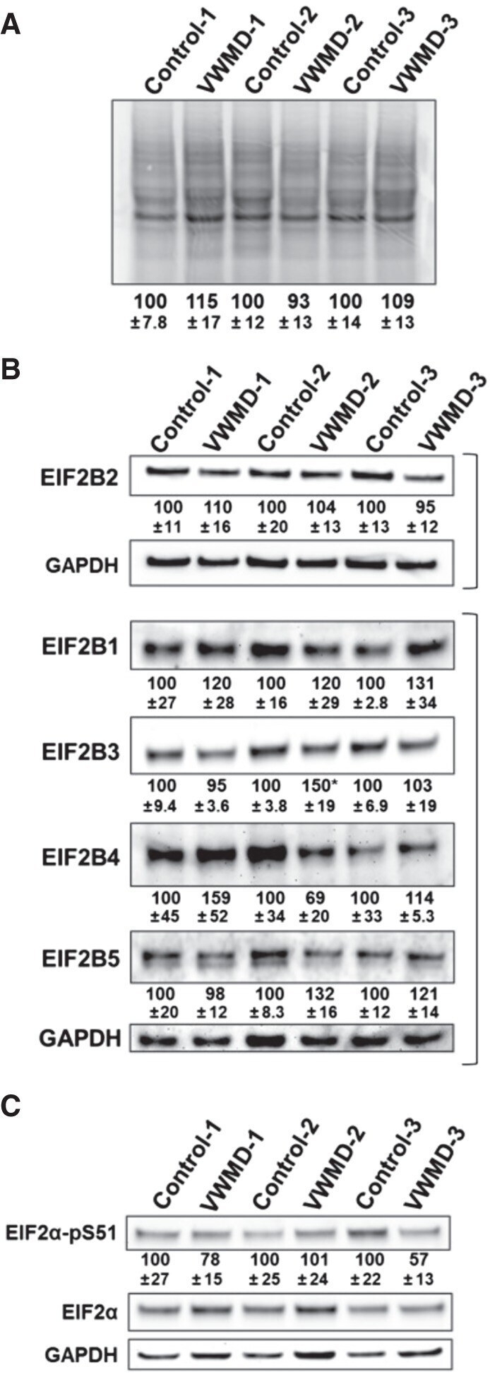
Submitted references EIF2B2 mutations in vanishing white matter disease hypersuppress translation and delay recovery during the integrated stress response.
Moon SL, Parker R
RNA (New York, N.Y.) 2018 Jun;24(6):841-852
RNA (New York, N.Y.) 2018 Jun;24(6):841-852
No comments: Submit comment
Supportive validation
- Submitted by
- Invitrogen Antibodies (provider)
- Main image

- Experimental details
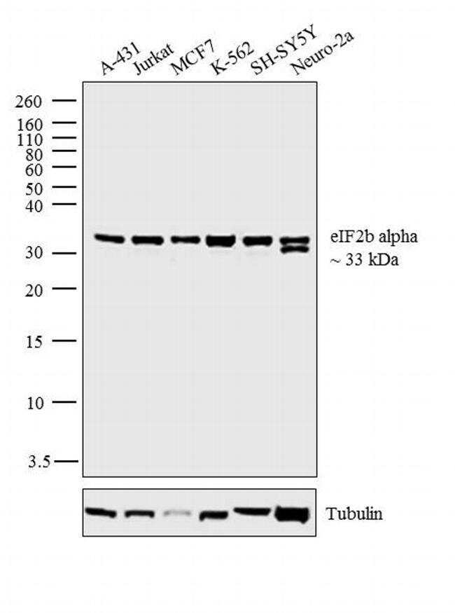
- Western blot analysis was performed on whole cell extracts (30 µg lysate) of A-431 (Lane 1), Jurkat (Lane 2), MCF7 (Lane 3), K-562 (Lane 4), SH-SY5Y (Lane 5) and Neuro-2a (Lane 6). The blot was probed with Anti-eIF2b alpha Polyclonal Antibody (Product # PA5-28992, 1:5,000 dilution) and detected by chemiluminescence using Goat anti-Rabbit IgG (Heavy Chain) Superclonal™ Secondary Antibody, HRP conjugate (Product # A27036, 0.25 µg/mL, 1:4,000 dilution). A 33 kDa band corresponding to eIF2b alpha was observed across the cell lines tested.
- Submitted by
- Invitrogen Antibodies (provider)
- Main image

- Experimental details
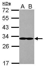
- Western Blot using eIF2b alpha Polyclonal Antibody (Product # PA5-28992). Sample (30 µg of whole cell lysate). Lane A: A431. Lane B: Jurkat. 12% SDS PAGE. eIF2b alpha Polyclonal Antibody (Product # PA5-28992) diluted at 1:10,000.
- Submitted by
- Invitrogen Antibodies (provider)
- Main image
- Experimental details
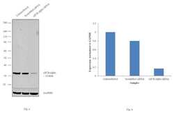
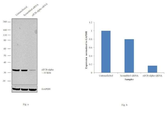
- Knockdown of EIF2B alpha was achieved by transfecting A431 with EIF2B alpha specific siRNAs (Silencer® select Product # s4558, s4559). Western blot analysis (Fig. a) was performed using membrane enriched extracts from the EIF2B alpha knockdown cells (lane 3), non-specific scrambled siRNA transfected cells (lane 2) and untransfected cells (lane 1). The blot was probed with EIF2B alpha Polyclonal Antibody (Product # PA5-28992, 1:5,000 dilution) and Goat anti-Rabbit IgG (Heavy Chain) Superclonal™ Secondary Antibody, HRP conjugate (Product # A27036, 0.25 µg/mL, 1:4,000 dilution). Densitometric analysis of this western blot is shown in histogram (Fig. b). Decrease in signal upon siRNA mediated knock down confirms that antibody is specific to EIF2B alpha.
Supportive validation
- Submitted by
- Invitrogen Antibodies (provider)
- Main image
- Experimental details
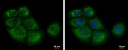
- eIF2b alpha Polyclonal Antibody detects EIF2B1 protein at cytoplasm by immunofluorescent analysis. Sample: A431 cells were fixed in 4% paraformaldehyde at RT for 15 min. Green: EIF2B1 protein stained by eIF2b alpha Polyclonal Antibody (Product # PA5-28992) diluted at 1:500. Blue: Hoechst 33342 staining.

- Submitted by
- Invitrogen Antibodies (provider)
- Main image

- Experimental details
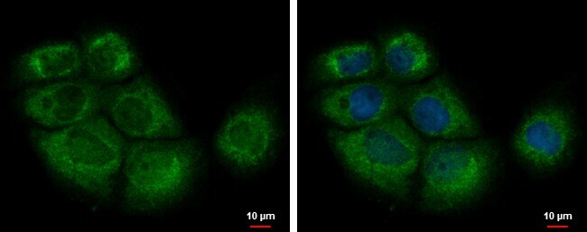
- eIF2b alpha Polyclonal Antibody detects EIF2B1 protein at cytoplasm by immunofluorescent analysis. Sample: A431 cells were fixed in 4% paraformaldehyde at RT for 15 min. Green: EIF2B1 protein stained by eIF2b alpha Polyclonal Antibody (Product # PA5-28992) diluted at 1:500. Blue: Hoechst 33342 staining.
Supportive validation
- Submitted by
- Invitrogen Antibodies (provider)
- Main image
- Experimental details
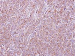
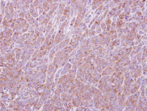
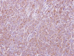
- Immunohistochemical analysis of paraffin-embedded BT483 xenograft, using EIF2B1 (Product # PA5-28992) antibody at 1:500 dilution. Antigen Retrieval: EDTA based buffer, pH 8.0, 15 min.
- Submitted by
- Invitrogen Antibodies (provider)
- Main image
- Experimental details
- Immunohistochemical analysis of paraffin-embedded BT483 xenograft, using EIF2B1 (Product # PA5-28992) antibody at 1:500 dilution. Antigen Retrieval: EDTA based buffer, pH 8.0, 15 min.

Supportive validation
- Submitted by
- Invitrogen Antibodies (provider)
- Main image
- Experimental details
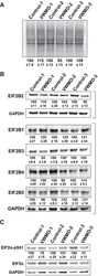
- FIGURE 1. VWMD cell lines with EIF2B2 mutations have normal translation activity in unstressed conditions. ( A ) Equal numbers of control and VWMD patient cell lines were fed 35 S-labeled met and cys, lysed and proteins separated on 4%-12% NuPAGE SDS PAGE gels and detected by phosphorimaging. The intensity of each lane was quantified and the averages +-SD of the lane intensity of each VWMD patient sample relative to the matched control from three independent experiments is shown below a representative gel. ( B ) The levels of EIF2B1, EIF2B2, EIF2B3, EIF2B4, and EIF2B5 proteins in VWMD patient and control lymphoblasts was assessed by western blotting. Brackets enclose blots for which the indicated EIF2B protein was stripped and reprobed for GAPDH as a loading control. Quantification shows the average relative EIF2B protein levels from three independent experiments +-SEM below a representative blot. ( C ) Phospho-eIF2alpha levels were determined by western blotting in VWMD patient and matched controls. The average abundance of phospho-eIF2alpha (""EIF2alpha-pS51"") relative to total EIF2alpha from three independent experiments +-SEM is shown below a representative blot. Student's t -test was used to assess significance in VWMD samples versus control for all experiments.