Explore
 Validate
 Learn
Learn Western blot
Antibody data
- Antibody Data
- Antigen structure
- References [2]
- Comments [0]
- Validations
- Western blot [2]
- Immunocytochemistry [1]
- Immunohistochemistry [3]
- Other assay [1]
Submit
Validation data
Reference
Comment
Report error
- Product number
- PA5-73015 - Provider product page

- Provider
- Invitrogen Antibodies
- Product name
- SLUG Polyclonal Antibody
- Antibody type
- Polyclonal
- Antigen
- Other
- Reactivity
- Human, Mouse, Canine
- Host
- Rabbit
- Isotype
- IgG
- Vial size
- 100 µg
- Concentration
- 1 mg/mL
- Storage
- Store at 4°C short term. For long term storage, store at -20°C, avoiding freeze/thaw cycles.
Submitted references Bile acids increase intestinal marker expression via the FXR/SNAI2/miR-1 axis in the stomach.
The metastasis suppressor, NDRG1, attenuates oncogenic TGF-β and NF-κB signaling to enhance membrane E-cadherin expression in pancreatic cancer cells.
Wang N, Wu S, Zhao J, Chen M, Zeng J, Lu G, Wang J, Zhang J, Liu J, Shi Y
Cellular oncology (Dordrecht) 2021 Oct;44(5):1119-1131
Cellular oncology (Dordrecht) 2021 Oct;44(5):1119-1131
The metastasis suppressor, NDRG1, attenuates oncogenic TGF-β and NF-κB signaling to enhance membrane E-cadherin expression in pancreatic cancer cells.
Menezes SV, Fouani L, Huang MLH, Geleta B, Maleki S, Richardson A, Richardson DR, Kovacevic Z
Carcinogenesis 2019 Jul 6;40(6):805-818
Carcinogenesis 2019 Jul 6;40(6):805-818
No comments: Submit comment
Supportive validation
- Submitted by
- Invitrogen Antibodies (provider)
- Main image

- Experimental details
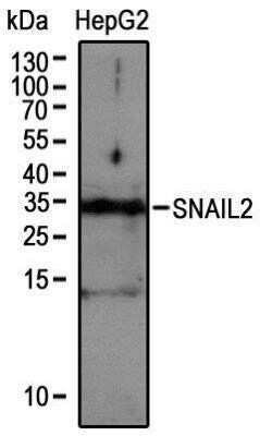
- Western blot analysis of SLUG in extracts from HepG2 cells. Samples were incubated in SLUG polyclonal antibody (Product # PA5-73015) using a dilution of 1:1000.
- Submitted by
- Invitrogen Antibodies (provider)
- Main image
- Experimental details
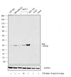
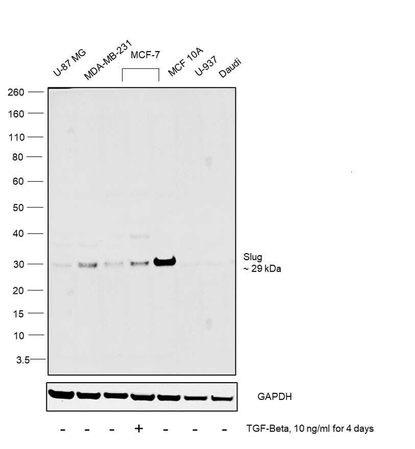
- Western blot was performed using Anti-SLUG Polyclonal Antibody (Product # PA5-73015) and a 29kDa band corresponding to Slug was observed across all the cell lines tested and increased upon TGF-Beta treatment in MCF7. Whole cell extracts (30 µg lysate) of U-87 MG (Lane 1), MDA-MB-231 (Lane 2), MCF-7 (Lane 3), MCF-7 treated with TGF-Beta (10ng/ml for 4 days) (Lane 4), MCF 10A (Lane 5), U-937 (Lane 6) and Daudi were electrophoresed using NuPAGE™ 4-12% Bis-Tris Protein Gel (Product # NP0322BOX). Resolved proteins were then transferred onto a nitrocellulose membrane (Product # IB23001) by iBlot® 2 Dry Blotting System (Product # IB21001). The blot was probed with the primary antibody (1:2000 dilution) and detected by chemiluminescence with Goat anti-Rabbit IgG (H+L) Superclonal™ Recombinant Secondary Antibody, HRP (Product # A27036, 1:4000 dilution) using the iBright FL 1000 (Product # A32752). Chemiluminescent detection was performed using Novex® ECL Chemiluminescent Substrate Reagent Kit (Product # WP20005).
Supportive validation
- Submitted by
- Invitrogen Antibodies (provider)
- Main image
- Experimental details
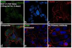
- Immunofluorescence analysis of SLUG was performed using 70% confluent log phase MCF-7 cells treated with 10 ng/mL TGF-Beta for 4 days. The cells were fixed with 4% Paraformaldehyde for 10 minutes, permeabilized with 0.1% Triton™ X-100 for 10 minutes, and blocked with 2% BSA for 10 minutes at room temperature. The cells were labeled Anti-SLUG Polyclonal Antibody (Product # Anti-SLUG Polyclonal Antibody (Product # PA5-73015) at 1:100 dilution in 0.1% BSA, incubated at 4 degree celsius overnight and then labeled with Goat anti-Rabbit IgG (H+L) Superclonal™ Recombinant Secondary Antibody, Alexa Fluor® 488 (Product # A27034) for 45 minutes at room temperature (Panel a: Green). Nuclei (Panel b: Blue) were stained with SlowFade® Gold Antifade Mountant with DAPI (Product # S36938). F-actin (Panel c: Red) was stained with Rhodamine Phalloidin (Product # R415, 1:300). Panel d represents the merged image showing nuclear localization. Panel e represents untreated MCF-7 cells having low expression of SLUG. Panel f represents control cells with no primary antibody to assess background. The images were captured at 60X magnification.

Supportive validation
- Submitted by
- Invitrogen Antibodies (provider)
- Main image

- Experimental details
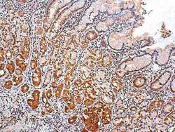
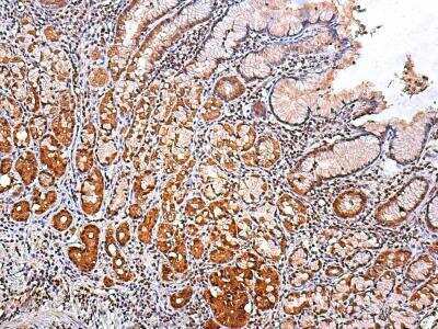
- Immunohistochemical analysis of SLUG in gastric cancer. Samples were incubated in SLUG polyclonal antibody (Product # PA5-73015) using a dilution of 1:25. Image objective 400X.

- Submitted by
- Invitrogen Antibodies (provider)
- Main image
- Experimental details
- Immunohistochemical analysis of SLUG in gastric cancer. Samples were incubated in SLUG polyclonal antibody (Product # PA5-73015) using a dilution of 1:25. Image objective 100X.
- Submitted by
- Invitrogen Antibodies (provider)
- Main image
- Experimental details
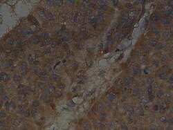
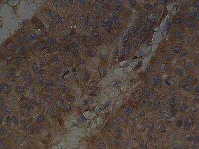
- Immunohistochemical analysis of SLUG in liver cancer. Samples were incubated in SLUG polyclonal antibody (Product # PA5-73015) using a dilution of 1:25. Image objective 400X.
Supportive validation
- Submitted by
- Invitrogen Antibodies (provider)
- Main image
- Experimental details
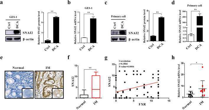
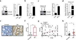
- Fig. 3 Overexpression of SNAI2 in IM cells. (a) GES-1 cells were treated as in Fig. 1b . The protein level of SNAI2 was detected by Western blotting (Left) and the results were normalized to beta-actin (Right). (b) SNAI2 mRNA levels examined in GES-1 cells after DCA treatment by qRT-PCR. (c, d) Primary cultured cells were treated with DCA as for GES-1 cells, after which SNAI2 was detected at protein and mRNA levels, respectively. (e) Representative pictures of IHC staining for SNAI2 in normal and gastric IM tissues. Scale bars: 100 mum and 500 mum (insets). (f) Quantification of IHC staining for SNAI2. (g) Correlation between SNAI2 and FXR expression in IM specimens. (h) SNAI2 mRNA levels in 10 pairs of matched human IM/normal specimens. * p < 0.05; ** p < 0.01