Explore
 Validate
 Learn
Learn Western blot
 Immunohistochemistry
Antibody data
- Antibody Data
- Antigen structure
- References [1]
- Comments [0]
- Validations
- Immunohistochemistry [1]
- Other assay [1]
Submit
Validation data
Reference
Comment
Report error
- Product number
- PA5-31432 - Provider product page

- Provider
- Invitrogen Antibodies
- Product name
- ARF5 Polyclonal Antibody
- Antibody type
- Polyclonal
- Antigen
- Recombinant full-length protein
- Description
- Recommended positive controls: 293T, Raji, mouse brain. Predicted reactivity: Mouse (100%), Rat (100%), Zebrafish (95%), Xenopus laevis (85%), Pig (100%), Chicken (98%), Sheep (100%), Bovine (100%). Store product as a concentrated solution. Centrifuge briefly prior to opening the vial.
- Reactivity
- Human, Mouse
- Host
- Rabbit
- Isotype
- IgG
- Vial size
- 100 μL
- Concentration
- 0.43 mg/mL
- Storage
- Store at 4°C short term. For long term storage, store at -20°C, avoiding freeze/thaw cycles.
Submitted references Which Way In? The RalF Arf-GEF Orchestrates Rickettsia Host Cell Invasion.
Rennoll-Bankert KE, Rahman MS, Gillespie JJ, Guillotte ML, Kaur SJ, Lehman SS, Beier-Sexton M, Azad AF
PLoS pathogens 2015 Aug;11(8):e1005115
PLoS pathogens 2015 Aug;11(8):e1005115
No comments: Submit comment
Supportive validation
- Submitted by
- Invitrogen Antibodies (provider)
- Main image
- Experimental details
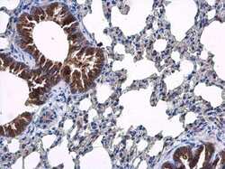
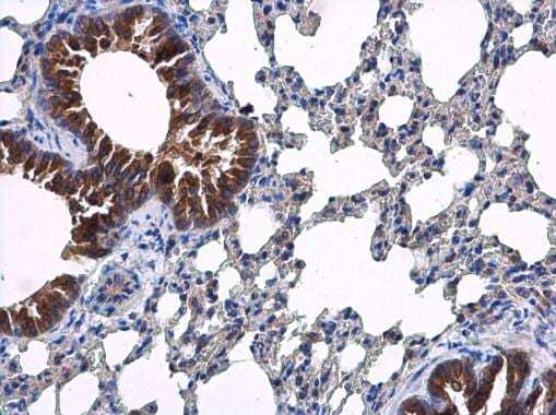
- Immunohistochemistry (Paraffin) analysis of ARF5 was performed in paraffin-embedded mouse lung tissue using ARF5 Polyclonal Antibody (Product # PA5-31432) at a dilution of 1:500.
Supportive validation
- Submitted by
- Invitrogen Antibodies (provider)
- Main image
- Experimental details
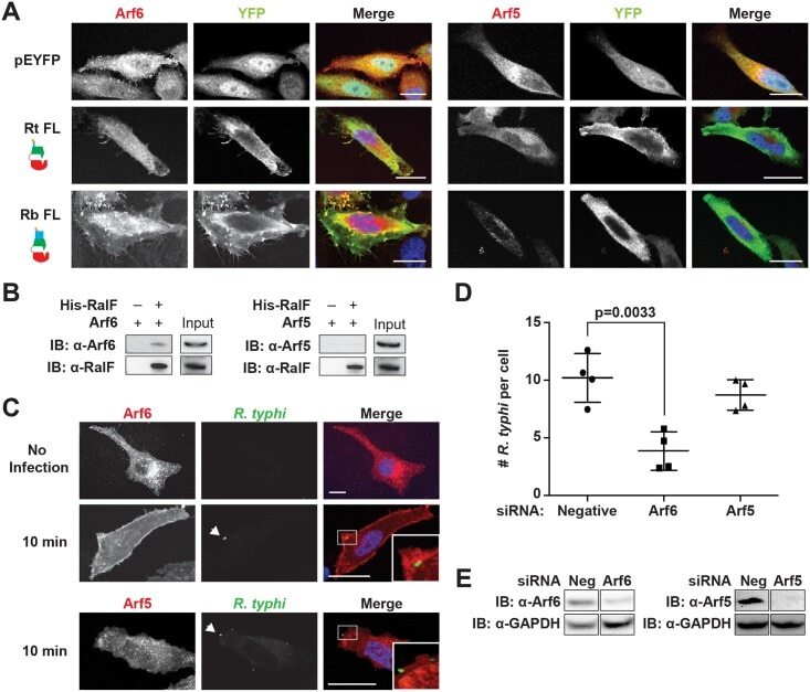
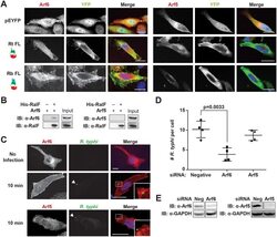
- Fig 6 Arf6 is recruited by R . typhi RalF and is required for infection. (A) Ectopically expressed RalF RtFL co-localizes with Arf6 but not Arf5. HeLa cells co-expressing EYFP, EYFP-RalF RtFL or EYFP-RalF RbFL and mRFP-Arf6 (left) or -Arf5 (right) were fixed with 4% para-formaldehyde. Nuclei were stained with DAPI (blue). (Scale bar: 10 mum). (B) RalF RtFL pull-down of Arf6. Lysates from HEK293T cells expressing mRFP-Arf5 or -Arf6 were incubated with HisPur Cobalt resin bound with rHis-RalF RtFL or resin alone. Bound proteins were eluted with imidazole and analyzed by protein immunoblot using antibodies as indicated. (C) Arf6 is recruited during R . typhi entry. HeLa cells expressing mRFP-Arf5 or -Arf6 (red) were infected with partially purified R . typhi (MOI ~100). Ten minutes post infection, cells were fixed and R . typhi detected with anti- R . typhi serum (green). DAPI (blue) is shown in the merged image. Boxed regions are enlarged to show detail. White arrowheads indicate R . typhi . (Scale bar: 5 mum). (D) Arf6 knockdown inhibits R . typhi infection. HeLa cells transfected with negative, Arf6, or Arf5 siRNA were infected with partially purified R . typhi (MOI ~100). At 2 hrs post infection, cells were fixed, plasma membrane stained with Alexa Fluor 594 wheat germ agglutinin, and R . typhi detected with rat anti- R . typhi serum and Alexa Fluor 488 anti-rat antibody. The number of R . typhi per host cell was counted for 100 host cells for three independent experiments.