 Explore
 Validate
 Learn
Learn Western blot
 ELISA
ELISAAntibody data
- Antibody Data
- Antigen structure
- References [14]
- Comments [0]
- Validations
- Western blot [1]
- Immunocytochemistry [1]
- Immunohistochemistry [2]
Submit
Validation data
Reference
Comment
Report error
- Product number
- 15340-1-AP - Provider product page

- Provider
- Proteintech Group
- Proper citation
- Proteintech Cat#15340-1-AP, RRID:AB_2254051
- Product name
- RPL7A antibody
- Antibody type
- Polyclonal
- Description
- RPL7A antibody (Cat. #15340-1-AP) is a rabbit polyclonal antibody that shows reactivity with human, mouse, rat and has been validated for the following applications: IF, IHC, IP, WB,ELISA.
- Reactivity
- Human, Mouse, Rat
- Host
- Rabbit
- Conjugate
- Unconjugated
- Isotype
- IgG
- Vial size
- 20ul, 150ul
Submitted references The RNA binding protein Arid5a is an activator of TNF signaling in rheumatoid arthritis.
RiboBright reveals cell-type-specific differences in ribosome organization and movement.
Decoding the Molecular Landscape of Prepubertal Oocyte Maturation: GTPBP4 as a Key Driver of In Vitro Developmental Competence.
DDX41 resolves G-quadruplexes to maintain erythroid genome integrity and prevent cGAS-mediated cell death.
The RNA binding protein Arid5a drives IL-17-dependent autoantibody-induced glomerulonephritis.
DDX41 dissolves G-quadruplexes to maintain erythroid genome integrity and prevent cGAS-mediated cell death.
LLPS of FXR proteins drives replication organelle clustering for β-coronaviral proliferation.
Nuclear and cytoplasmic specific RNA binding proteome enrichment and its changes upon ferroptosis induction.
UBE2S targets RPL26 for ubiquitination and degradation to promote non-small cell lung cancer progression via regulating c-Myc.
The induction of p53 correlates with defects in the production, but not the levels, of the small ribosomal subunit and stalled large ribosomal subunit biogenesis.
Inefficient quality control of ribosome stalling during APP synthesis generates CAT-tailed species that precipitate hallmarks of Alzheimer's disease.
Estrogen-Related Hormones Induce Apoptosis by Stabilizing Schlafen-12 Protein Turnover.
Phosphorylation-dependent Regnase-1 release from endoplasmic reticulum is critical in IL-17 response.
Regnase-1 and Roquin Regulate a Common Element in Inflammatory mRNAs by Spatiotemporally Distinct Mechanisms.
Li Y, Dey I, Vyas SP, Synackova A, Li D, Lubberts E, Ascherman DP, Draber P, Gaffen SL
JCI insight 2026 Jan 23;11(2)
JCI insight 2026 Jan 23;11(2)
RiboBright reveals cell-type-specific differences in ribosome organization and movement.
Poulladofonou G, Grandi C, Hu X, Yesley P, Velema WA, Hansen MMK
Nature communications 2026 Feb 13;17(1)
Nature communications 2026 Feb 13;17(1)
Decoding the Molecular Landscape of Prepubertal Oocyte Maturation: GTPBP4 as a Key Driver of In Vitro Developmental Competence.
Qin J, Wei Y, Ning A, Hu W, Wan P, Cao B, Pan B, Lv T, Du K, Yao X, Zou S, Chen X, Zang S, Ye J, Yu G, Liang Q, Shen L, Zhang L, Chen X, Cheng K, Meng L, Zhou G
Cell proliferation 2025 Nov;58(11):e70017
Cell proliferation 2025 Nov;58(11):e70017
DDX41 resolves G-quadruplexes to maintain erythroid genome integrity and prevent cGAS-mediated cell death.
Bi H, Ren K, Wang P, Li E, Han X, Wang W, Yang J, Aydemir I, Tao K, Ma R, Godley LA, Liu Y, Shukla V, Bartom ET, Tang Y, Blanc L, Sukhanova M, Ji P
Nature communications 2025 Aug 5;16(1):7195
Nature communications 2025 Aug 5;16(1):7195
The RNA binding protein Arid5a drives IL-17-dependent autoantibody-induced glomerulonephritis.
Li Y, Vyas SP, Mehta I, Asada N, Dey I, Taylor TC, Bechara R, Amatya N, Aggor FEY, Coleman BM, Li DD, Yamamoto K, Ezenwa O, Sun Y, Sterneck E, McManus CJ, Panzer U, Biswas PS, Savan R, Das J, Gaffen SL
The Journal of experimental medicine 2024 Sep 2;221(9)
The Journal of experimental medicine 2024 Sep 2;221(9)
DDX41 dissolves G-quadruplexes to maintain erythroid genome integrity and prevent cGAS-mediated cell death.
Bi H, Ren K, Wang P, Li E, Han X, Wang W, Yang J, Aydemir I, Tao K, Godley L, Liu Y, Shukla V, Bartom ET, Tang Y, Blanc L, Sukhanova M, Ji P
bioRxiv : the preprint server for biology 2024 Oct 17;
bioRxiv : the preprint server for biology 2024 Oct 17;
LLPS of FXR proteins drives replication organelle clustering for β-coronaviral proliferation.
Li M, Hou Y, Zhou Y, Yang Z, Zhao H, Jian T, Yu Q, Zeng F, Liu X, Zhang Z, Zhao YG
The Journal of cell biology 2024 Jun 3;223(6)
The Journal of cell biology 2024 Jun 3;223(6)
Nuclear and cytoplasmic specific RNA binding proteome enrichment and its changes upon ferroptosis induction.
Sun H, Fu B, Qian X, Xu P, Qin W
Nature communications 2024 Jan 29;15(1):852
Nature communications 2024 Jan 29;15(1):852
UBE2S targets RPL26 for ubiquitination and degradation to promote non-small cell lung cancer progression via regulating c-Myc.
Gong D, Rao X, Min Z, Liu X, Xin H, Zhou P, Yang L, Li D
American journal of cancer research 2023;13(8):3705-3720
American journal of cancer research 2023;13(8):3705-3720
The induction of p53 correlates with defects in the production, but not the levels, of the small ribosomal subunit and stalled large ribosomal subunit biogenesis.
Eastham MJ, Pelava A, Wells GR, Lee JK, Lawrence IR, Stewart J, Deichner M, Hertle R, Watkins NJ, Schneider C
Nucleic acids research 2023 Sep 22;51(17):9397-9414
Nucleic acids research 2023 Sep 22;51(17):9397-9414
Inefficient quality control of ribosome stalling during APP synthesis generates CAT-tailed species that precipitate hallmarks of Alzheimer's disease.
Rimal S, Li Y, Vartak R, Geng J, Tantray I, Li S, Huh S, Vogel H, Glabe C, Grinberg LT, Spina S, Seeley WW, Guo S, Lu B
Acta neuropathologica communications 2021 Oct 18;9(1):169
Acta neuropathologica communications 2021 Oct 18;9(1):169
Estrogen-Related Hormones Induce Apoptosis by Stabilizing Schlafen-12 Protein Turnover.
Li D, Chen J, Ai Y, Gu X, Li L, Che D, Jiang Z, Li L, Chen S, Huang H, Wang J, Cai T, Cao Y, Qi X, Wang X
Molecular cell 2019 Sep 19;75(6):1103-1116.e9
Molecular cell 2019 Sep 19;75(6):1103-1116.e9
Phosphorylation-dependent Regnase-1 release from endoplasmic reticulum is critical in IL-17 response.
Tanaka H, Arima Y, Kamimura D, Tanaka Y, Takahashi N, Uehata T, Maeda K, Satoh T, Murakami M, Akira S
The Journal of experimental medicine 2019 Jun 3;216(6):1431-1449
The Journal of experimental medicine 2019 Jun 3;216(6):1431-1449
Regnase-1 and Roquin Regulate a Common Element in Inflammatory mRNAs by Spatiotemporally Distinct Mechanisms.
Mino T, Murakawa Y, Fukao A, Vandenbon A, Wessels HH, Ori D, Uehata T, Tartey S, Akira S, Suzuki Y, Vinuesa CG, Ohler U, Standley DM, Landthaler M, Fujiwara T, Takeuchi O
Cell 2015 May 21;161(5):1058-1073
Cell 2015 May 21;161(5):1058-1073
No comments: Submit comment
Supportive validation
- Submitted by
- Proteintech Group (provider)
- Main image

- Experimental details
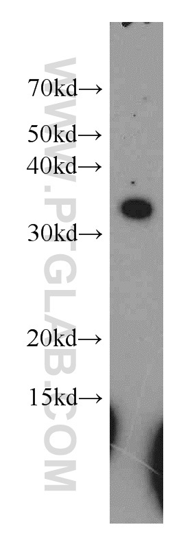
- MCF7 cells were subjected to SDS PAGE followed by western blot with 15340-1-AP(RPL7A antibody) at dilution of 1:500
- Sample type
- cell line
Supportive validation
- Submitted by
- Proteintech Group (provider)
- Main image

- Experimental details
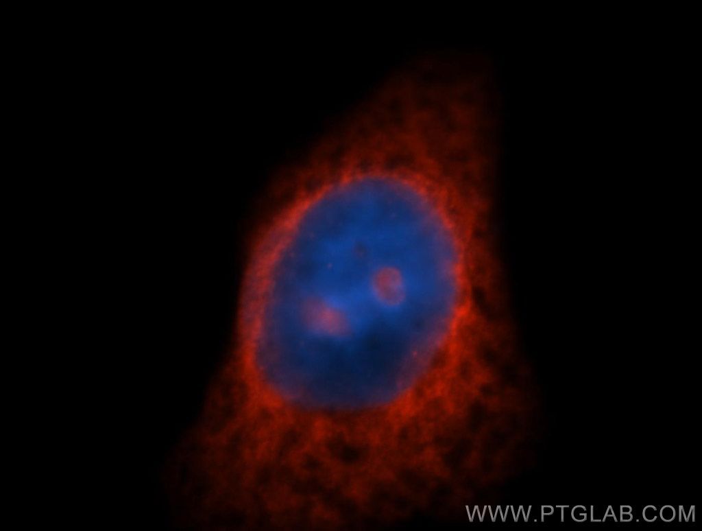
- Immunofluorescent analysis of HepG2 cells, using RPL7A antibody 15340-1-AP at 1:50 dilution and Rhodamine-labeled goat anti-rabbit IgG (red). Blue pseudocolor = DAPI (fluorescent DNA dye).
- Sample type
- cell line
Supportive validation
- Submitted by
- Proteintech Group (provider)
- Main image

- Experimental details
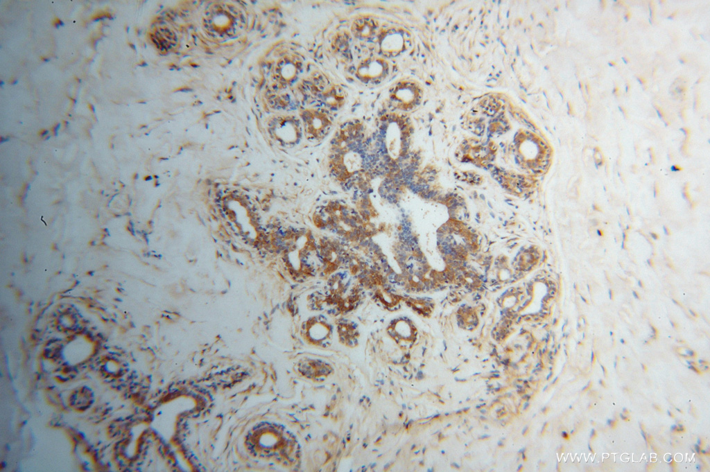
- Immunohistochemical of paraffin-embedded human breast cancer using 15340-1-AP(RPL7A antibody) at dilution of 1:100 (under 10x lens)
- Sample type
- tissue
- Submitted by
- Proteintech Group (provider)
- Main image
- Experimental details
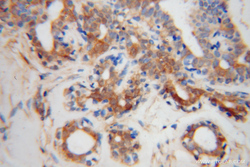
- Immunohistochemical of paraffin-embedded human breast cancer using 15340-1-AP(RPL7A antibody) at dilution of 1:100 (under 40x lens)
- Sample type
- tissue
