Explore
 Validate
 Learn
Learn Western blot
 Immunocytochemistry
Antibody data
- Antibody Data
- Antigen structure
- References [1]
- Comments [0]
- Validations
- Immunocytochemistry [2]
- Immunohistochemistry [5]
Submit
Validation data
Reference
Comment
Report error
- Product number
- PA5-61289 - Provider product page

- Provider
- Invitrogen Antibodies
- Product name
- SUCLG2 Polyclonal Antibody
- Antibody type
- Polyclonal
- Antigen
- Recombinant protein fragment
- Description
- Immunogen sequence: FGGIVNCAII ANGITKACRE LELKVPLVVR LEGTNVQEAQ KILNNSGLPI TSAIDLEDAA KKAVASVAKK Highest antigen sequence identity to the following orthologs: Mouse - 94%, Rat - 93%.
- Reactivity
- Human
- Host
- Rabbit
- Isotype
- IgG
- Vial size
- 100 μL
- Concentration
- 0.6 mg/mL
- Storage
- Store at 4°C short term. For long term storage, store at -20°C, avoiding freeze/thaw cycles.
Submitted references Mesothelial Cell HIF1α Expression Is Metabolically Downregulated by Metformin to Prevent Oncogenic Tumor-Stromal Crosstalk.
Hart PC, Kenny HA, Grassl N, Watters KM, Litchfield LM, Coscia F, Blaženović I, Ploetzky L, Fiehn O, Mann M, Lengyel E, Romero IL
Cell reports 2019 Dec 17;29(12):4086-4098.e6
Cell reports 2019 Dec 17;29(12):4086-4098.e6
No comments: Submit comment
Supportive validation
- Submitted by
- Invitrogen Antibodies (provider)
- Main image
- Experimental details
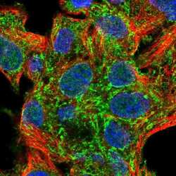
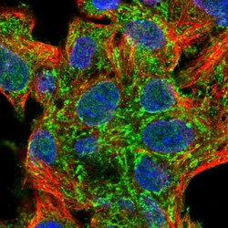
- Immunofluorescent staining of SUCLG2 in human cell line Hep G2 shows positivity in mitochondria. Samples were probed using a SUCLG2 Polyclonal Antibody (Product # PA5-61289).

- Submitted by
- Invitrogen Antibodies (provider)
- Main image
- Experimental details
- Immunofluorecent analysis of SUCLG2 in human cell line Hep G2 using SUCLG2 Polyclonal Antibody (Product # PA5-61289). Staining shows localization to mitochondria.

Supportive validation
- Submitted by
- Invitrogen Antibodies (provider)
- Main image

- Experimental details
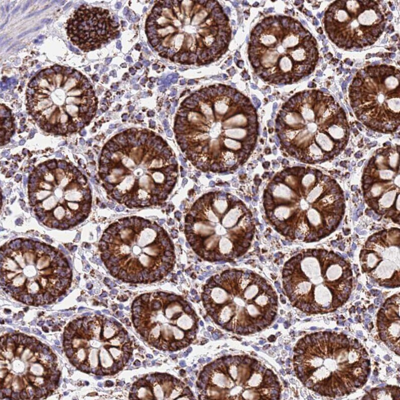
- Immunohistochemical staining of SUCLG2 in human colon using SUCLG2 Polyclonal Antibody (Product # PA5-61289) shows strong positivity in mitochondria in glandular cells.
- Submitted by
- Invitrogen Antibodies (provider)
- Main image
- Experimental details
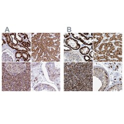
- Immunohistochemical staining of SUCLG2 in human kidney, liver, pancreas and testis using SUCLG2 Polyclonal Antibody (Product # PA5-61289) (A) shows similar protein distribution across tissues to an independent SUCLG2 Polyclonal Antibody (B).

- Submitted by
- Invitrogen Antibodies (provider)
- Main image
- Experimental details
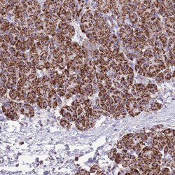
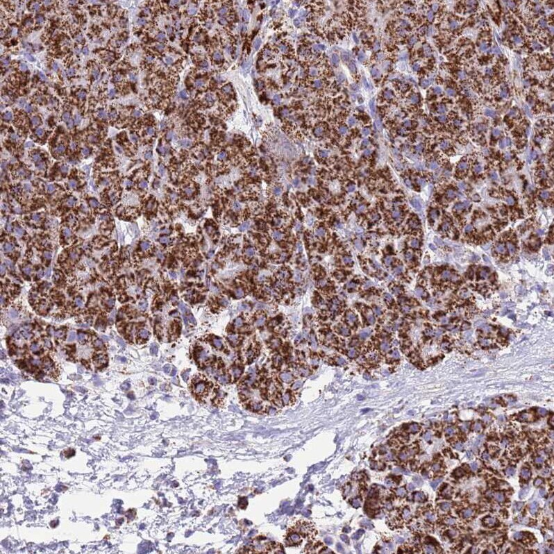
- Immunohistochemical staining of SUCLG2 in human pancreas using SUCLG2 Polyclonal Antibody (Product # PA5-61289) shows weak to moderate positivity in mitochondria in exocrine glandular cells.
- Submitted by
- Invitrogen Antibodies (provider)
- Main image

- Experimental details
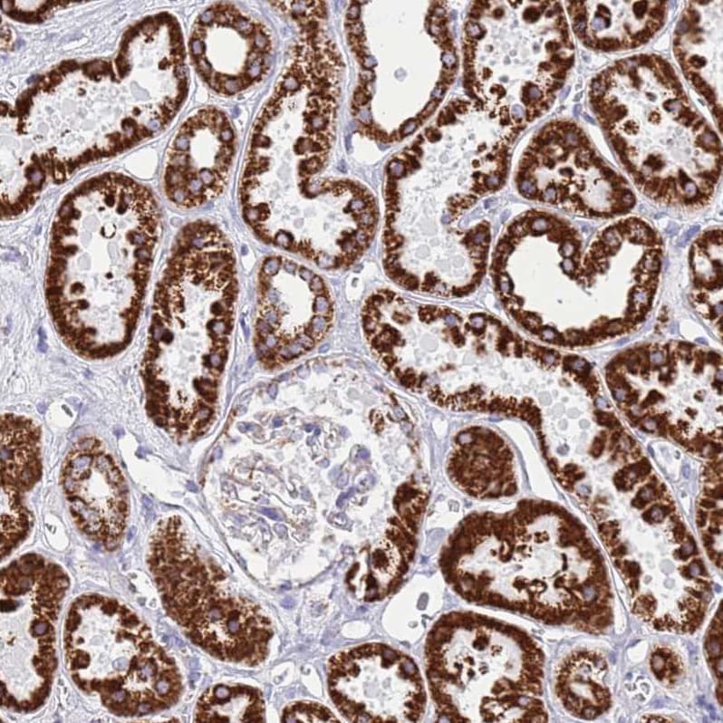
- Immunohistochemical staining of SUCLG2 in human kidney using SUCLG2 Polyclonal Antibody (Product # PA5-61289) shows strong positivity in mitochondria in cells in tubules.
- Submitted by
- Invitrogen Antibodies (provider)
- Main image

- Experimental details
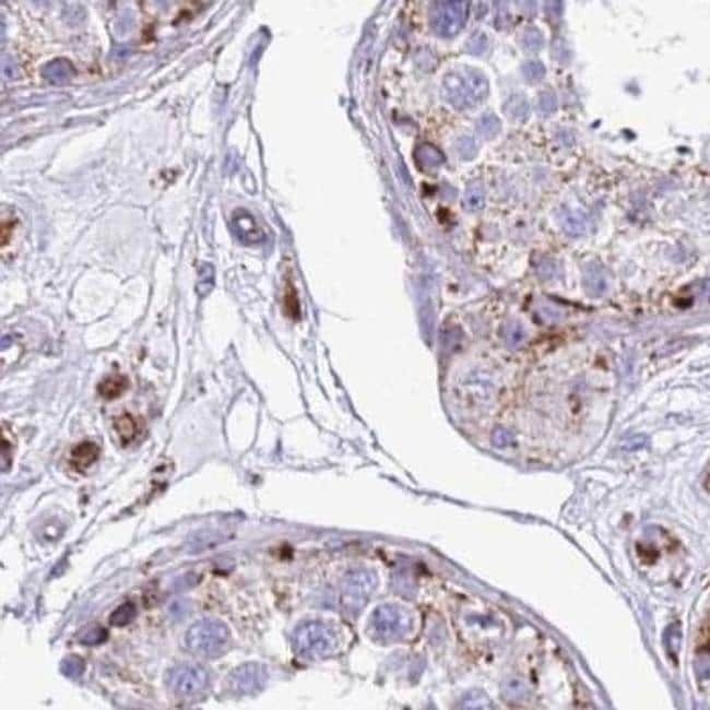
- Immunohistochemical staining of SUCLG2 in human testis using SUCLG2 Polyclonal Antibody (Product # PA5-61289).