 Explore
 Validate
 Learn
Learn Western blot
 ELISA
ELISAAntibody data
- Antibody Data
- Antigen structure
- References [2]
- Comments [0]
- Validations
- Western blot [2]
- Immunohistochemistry [1]
Submit
Validation data
Reference
Comment
Report error
- Product number
- NBP1-28605 - Provider product page

- Provider
- Novus Biologicals
- Proper citation
- Novus Cat#NBP1-28605, RRID:AB_1853360
- Product name
- Mouse Monoclonal MAT2A Antibody
- Antibody type
- Monoclonal
- Description
- Protein G purified.
- Reactivity
- Human
- Host
- Mouse
- Isotype
- IgG
- Vial size
- 0.1 ml
- Concentration
- 1.0 mg/ml
- Storage
- Store at 4C short term. Aliquot and store at -20C long term. Avoid freeze-thaw cycles.
Submitted references Induction of methionine adenosyltransferase 2A in tamoxifen-resistant breast cancer cells.
High-throughput screening for native autoantigen-autoantibody complexes using antibody microarrays.
Phuong NT, Kim SK, Im JH, Yang JW, Choi MC, Lim SC, Lee KY, Kim YM, Yoon JH, Kang KW
Oncotarget 2016 Mar 22;7(12):13902-16
Oncotarget 2016 Mar 22;7(12):13902-16
High-throughput screening for native autoantigen-autoantibody complexes using antibody microarrays.
Rho JH, Lampe PD
Journal of proteome research 2013 May 3;12(5):2311-20
Journal of proteome research 2013 May 3;12(5):2311-20
No comments: Submit comment
Supportive validation
- Submitted by
- Novus Biologicals (provider)
- Main image
- Experimental details
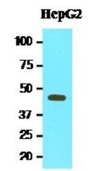
- Western Blot: MAT2A Antibody (3A2) [NBP1-28605] - Cell lysates of HepG2 (20ug) were resolved by SDS-PAGE, transferred to NC membrane and probed with anti-human MAT2A (1:1000). Proteins were visualized using a goat anti-mouse secondary antibody conjugated to HRP and an ECL detection system.

- Submitted by
- Novus Biologicals (provider)
- Main image
- Experimental details
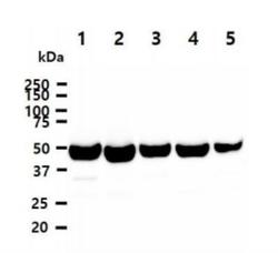
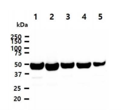
- Western Blot: MAT2A Antibody (3A2) [NBP1-28605] - The Cell lysates (40ug) were resolved by SDS-PAGE, transferred to PVDF membrane and probed with anti-human MAT2A antibody (1:1000). Proteins were visualized using a goat anti-mouse secondary antibody conjugated to HRP and an ECL detection system. Lane 1. : HeLa cell lysate Lane 2. : Ramos cell lysate Lane 3. : 293T cell lysate Lane 4. : Jurkat cell lysate Lane 5. : NIH-3T3 cell lysate
Supportive validation
- Submitted by
- Novus Biologicals (provider)
- Main image
- Experimental details
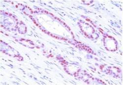
- Immunohistochemistry-Paraffin: MAT2A Antibody (3A2) [NBP1-28605] - Human MAT2A (1:50) for 2 hours at room temperature. Antigen retrieval was performed in 0.1M sodium citrate buffer and detected using Diaminobenzidine (DAB).
