Explore
 Validate
 Learn
LearnPB9502
antibody from Boster Biological Technology
Targeting: SCARB1
CD36L1, CLA-1, CLA1, SR-BI, SRB1
 Western blot
 Immunocytochemistry
Antibody data
- Antibody Data
- Antigen structure
- References [1]
- Comments [0]
- Validations
- Western blot [1]
Submit
Validation data
Reference
Comment
Report error
- Product number
- PB9502 - Provider product page

- Provider
- Boster Biological Technology
- Product name
- Anti-Scavenging Receptor SR-BI/SCARB1 Antibody Picoband™
- Antibody type
- Polyclonal
- Description
- Polyclonal antibody for SR BI/Scarb1 detection. Host: Rabbit.Size: 100μg/vial. Tested applications: Flow Cytometry. Reactive species: Mouse. SR BI/Scarb1 information: Molecular Weight: 56754 MW; Subcellular Localization: Cell membrane ; Multi-pass membrane protein . Membrane, caveola ; Multi-pass membrane protein . Predominantly localized to cholesterol and sphingomyelin-enriched domains within the plasma membrane, called caveolae; Tissue Specificity: Expressed primarily in liver and non-placental steroidogenic tissues.
- Reactivity
- Mouse, Rat
- Host
- Rabbit
- Vial size
- 100μg/vial
- Concentration
- Add 0.2ml of distilled water will yield a concentration of 500ug/ml.
- Storage
- At -20°C for one year. After reconstitution, at 4°C for one month. It can also be aliquoted and stored frozen at -20°C for a longer time. Avoid repeated freezing and thawing.
- Handling
- Add 0.2ml of distilled water will yield a concentration of 500ug/ml.
Submitted references Functional Deletion/Insertion Promoter Variants in SCARB1 Associated With Increased Susceptibility to Lipid Profile Abnormalities and Coronary Heart Disease.
Hu S, Hu D, Wei H, Li SY, Wang D, Li CZ, Jiang J, Wang D, Cui G, Wang D
Frontiers in cardiovascular medicine 2021;8:800873
Frontiers in cardiovascular medicine 2021;8:800873
No comments: Submit comment
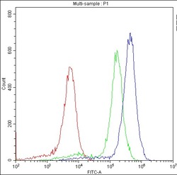
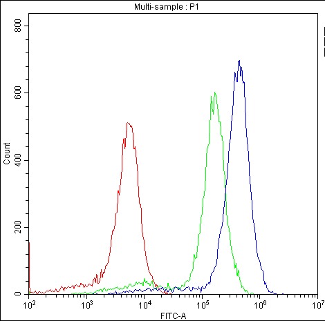
Supportive validation
- Submitted by
- Boster Biological Technology (provider)
- Main image
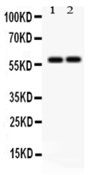
- Experimental details
- Western blot analysis of SCARB1 using anti-SCARB1 antibody (PB9502). Electrophoresis was performed on a 5-20% SDS-PAGE gel at 70V (Stacking gel) / 90V (Resolving gel) for 2-3 hours. The sample well of each lane was loaded with 50ug of sample under reducing conditions. Lane 1: Rat Testis Tissue Lysate, Lane 2: Mouse Testis Tissue Lysate. After Electrophoresis, proteins were transferred to a Nitrocellulose membrane at 150mA for 50-90 minutes. Blocked the membrane with 5% Non-fat Milk/ TBS for 1.5 hour at RT. The membrane was incubated with rabbit anti-SCARB1 antigen affinity purified polyclonal antibody (Catalog # PB9502) at 0.5 μg/mL overnight at 4°C, then washed with TBS-0.1%Tween 3 times with 5 minutes each and probed with a goat anti-rabbit IgG-HRP secondary antibody at a dilution of 1:10000 for 1.5 hour at RT. The signal is developed using an Enhanced Chemiluminescent detection (ECL) kit (Catalog # EK1002) with Tanon 5200 system. A specific band was detected for SCARB1 at approximately 57KD. The expected band size for SCARB1 is at 57KD.
- Additional image