 Explore
 Validate
 Learn
LearnPA1-16788
antibody from Invitrogen Antibodies
Targeting: SCARB1
CD36L1, CLA-1, CLA1, SR-BI, SRB1
 Western blot Immunocytochemistry
 Immunoprecipitation Immunohistochemistry Flow cytometry Other assay
Antibody data
- Antibody Data
- Antigen structure
- References [1]
- Comments [0]
- Validations
- Western blot [3]
- Immunocytochemistry [2]
- Immunohistochemistry [5]
- Flow cytometry [3]
- Other assay [2]
Submit
Validation data
Reference
Comment
Report error
- Product number
- PA1-16788 - Provider product page

- Provider
- Invitrogen Antibodies
- Product name
- SR-BI Polyclonal Antibody
- Antibody type
- Polyclonal
- Antigen
- Synthetic peptide
- Description
- Suggested positive control: antigen standard for SCARB1 (transient overexpression lysate).
- Reactivity
- Human, Mouse, Rat, Hamster, Porcine, Rabbit
- Host
- Rabbit
- Isotype
- IgG
- Vial size
- 100 μL
- Concentration
- 1 mg/mL
- Storage
- Store at 4°C short term. For long term storage, store at -20°C, avoiding freeze/thaw cycles.
Submitted references Insulin increases cholesterol uptake, lipid droplet content, and apolipoprotein B secretion in CaCo-2 cells by upregulating SR-BI via a PI3K, AKT, and mTOR-dependent pathway.
Fuentes M, Santander N, Cortés V
Journal of cellular biochemistry 2019 Feb;120(2):1550-1559
Journal of cellular biochemistry 2019 Feb;120(2):1550-1559
No comments: Submit comment
Supportive validation
- Submitted by
- Invitrogen Antibodies (provider)
- Main image
- Experimental details
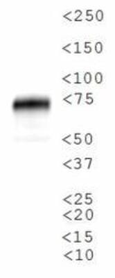
- Western blot analysis of SR-BI in human adrenal cell lysate. Sample was incubated in SR-BI polyclonal antibody (Product # PA1-16788).
- Submitted by
- Invitrogen Antibodies (provider)
- Main image

- Experimental details
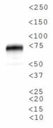
- Western blot analysis of SR-BI in 0.5 mg/mL HeLa lysate. Samples were incubated in SR-BI polyclonal antibody (Product # PA1-16788). This experiment was performed under reducing conditions using the 12-230 kDa separation system.

- Submitted by
- Invitrogen Antibodies (provider)
- Main image
- Experimental details
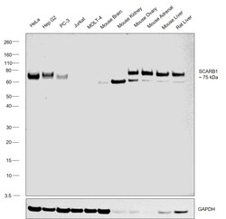
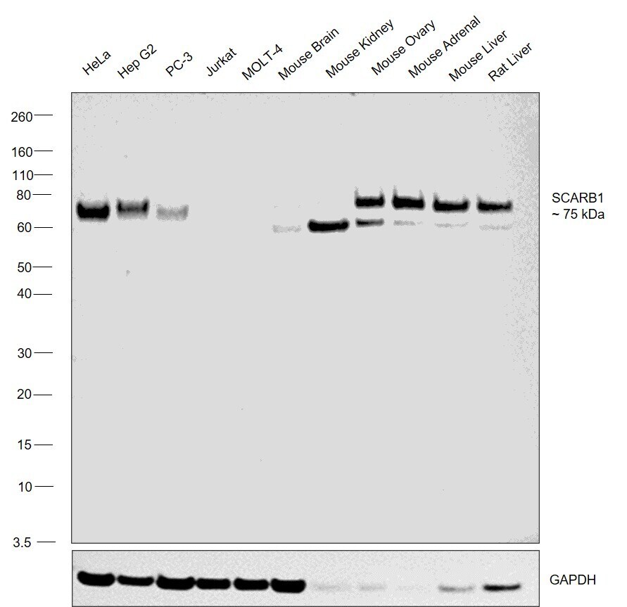
- Western blot was performed using Anti-SR-BI Polyclonal Antibody (Product # PA1-16788) and a 75 kDa band corresponding to SCARB1 was observed across across the cell lines and tissues tested. Membrane enriched extracts (30 µg lysate) of HeLa (Lane 1), Hep G2 (Lane 2), PC-3 (Lane 3), Jurkat (Lane 4), MOLT-4 (Lane 5), Mouse Brain (Lane 6), Mouse Kidney (Lane 7), Mouse Ovary (Lane 8), Mouse Adrenal Gland (Lane 9), Mouse Liver (Lane 10), Rat Liver (Lane 11) were electrophoresed using NuPAGE™ 4-12% Bis-Tris Protein Gel (Product # NP0322BOX). Resolved proteins were then transferred onto a nitrocellulose membrane (Product # IB23001) by iBlot® 2 Dry Blotting System (Product # IB21001). The blot was probed with the primary antibody (1:1000 dilution) and detected by chemiluminescence with Goat anti-Rabbit IgG (Heavy Chain) Superclonal™ Recombinant Secondary Antibody, HRP (Product # A27036,1:20000 dilution) using the iBright™ FL1500 Imaging System (Product # A44115). Chemiluminescent detection was performed using SuperSignal™ West Pico PLUS Chemiluminescent Substrate (Product # 34580).
Supportive validation
- Submitted by
- Invitrogen Antibodies (provider)
- Main image
- Experimental details
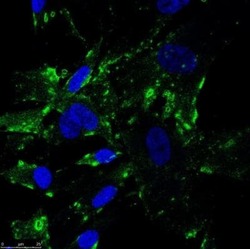
- Immunocytochemistry analysis of SR-BI in human fibroblast samples fixed in 4% PFA and permeabilized in PBS (0.2% Tween). Samples were incubated in SR-BI polyclonal antibody (Product # PA1-16788) using a dilution of 1:100 dilution in PBS (0.1% Tween) with 1% BSA followed by anti-rabbit conjugate to Alexa Fluor 488. Primary incubation overnight at 4 °C. SR-BI is shown in green and nucleus in blue (Hoescht 33342 stain).

- Submitted by
- Invitrogen Antibodies (provider)
- Main image

- Experimental details
- Immunocytochemistry analysis of SR-BI in human fibroblast samples fixed in 4% PFA and permeabilized in PBS (0.2% Tween). Samples were incubated in SR-BI polyclonal antibody (Product # PA1-16788) using a dilution of 1:100 dilution in PBS (0.1% Tween) with 1% BSA followed by anti-rabbit conjugate to Alexa Fluor 488. Primary incubation overnight at 4 °C. SR-BI is shown in green and nucleus in blue (Hoescht 33342 stain).

Supportive validation
- Submitted by
- Invitrogen Antibodies (provider)
- Main image
- Experimental details
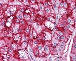
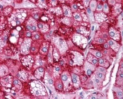
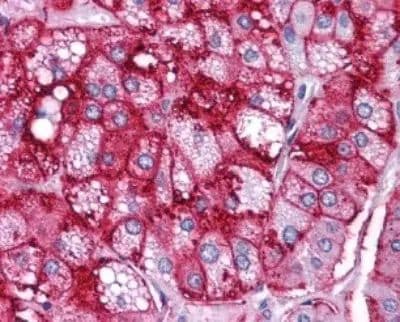
- Immunohistochemical analysis of SR-BI in human adrenal cortex. Samples were incubated in SR-BI polyclonal antibody (Product # PA1-16788).
- Submitted by
- Invitrogen Antibodies (provider)
- Main image

- Experimental details
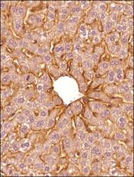
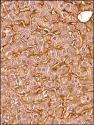
- Immunohistochemical analysis of SR-BI in formalin-fixed paraffin-embedded tissue section of mouse liver. Samples were incubated in SR-BI polyclonal antibody (Product # PA1-16788) using a dilution of 1:300 followed by HRP-DAB detection and hematoxylin counterstaining. The antibody generated mainly a membranous signal of SR-BI protein in the murine hepatocytes.

- Submitted by
- Invitrogen Antibodies (provider)
- Main image
- Experimental details
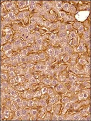
- Immunohistochemical analysis of SR-BI in formalin-fixed paraffin-embedded tissue section of mouse liver. Samples were incubated in SR-BI polyclonal antibody (Product # PA1-16788) using a dilution of 1:300 followed by HRP-DAB detection and hematoxylin counterstaining. The antibody generated a specific membrane signal of SR-BI protein in the murine hepatocytes.

- Submitted by
- Invitrogen Antibodies (provider)
- Main image
- Experimental details
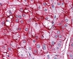
- Immunohistochemical analysis of SR-BI in human adrenal cortex. Samples were incubated in SR-BI polyclonal antibody (Product # PA1-16788).
- Submitted by
- Invitrogen Antibodies (provider)
- Main image
- Experimental details
- Immunohistochemical analysis of SR-BI in formalin-fixed paraffin-embedded tissue section of mouse liver. Samples were incubated in SR-BI polyclonal antibody (Product # PA1-16788) using a dilution of 1:300 followed by HRP-DAB detection and hematoxylin counterstaining. The antibody generated mainly a membranous signal of SR-BI protein in the murine hepatocytes.
Supportive validation
- Submitted by
- Invitrogen Antibodies (provider)
- Main image

- Experimental details
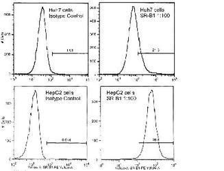
- Huh7 and HepG2 cells using SR-B1 antibody, Product # PA1-16788.
- Submitted by
- Invitrogen Antibodies (provider)
- Main image

- Experimental details
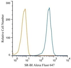
- Flow cytometry of SR-BI in HeLa cells (blue) and a matched isotype control (orange). Samples were incubated in SR-BI polyclonal antibody (Product # PA1-16788) using a dilution of 5 µg/mL for 30 minutes at room temperature. Cells were fixed with 4% PFA and then permeabilized with 0.1% saponin. Both antibodies were conjugated to Alexa Fluor 488.

- Submitted by
- Invitrogen Antibodies (provider)
- Main image
- Experimental details
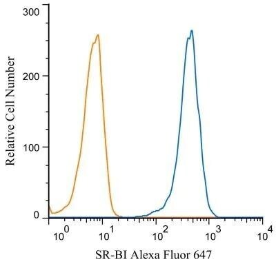
- Flow cytometry of SR-BI in HeLa cells (blue) and a matched isotype control (orange). Samples were incubated in SR-BI polyclonal antibody (Product # PA1-16788) using a dilution of 5 µg/mL for 30 minutes at room temperature. Cells were fixed with 4% PFA and then permeabilized with 0.1% saponin. Both antibodies were conjugated to Alexa Fluor 488.
Supportive validation
- Submitted by
- Invitrogen Antibodies (provider)
- Main image
- Experimental details
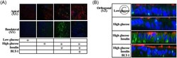
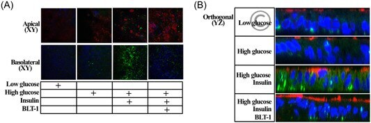
- 3 Insulin increases intracellular neutral lipid content in CaCo-2 cells by mechanisms that require SR-BI-mediated cholesterol uptake. A, CaCo-2 cells were incubated for 24 h with glucose at low (5 mM) or high (25 mM) concentrations, plus insulin (100 nM) and BLT-1 (1 muM) or vehicle and then subjected to confocal microscopy analysis to visualize SR-BI (red), lipid droplets, (green, BODIPY), and nuclei (blue, H33342), as indicated in Section 2. B, Digital orthogonal ( YZ ) projection reconstruction of confocal images of the cell cultures depicted in A, BODIPY, 4,4-difluoro-4-bora-3a,4a-diaza- s -indacene; BLT-1, block lipid transport-1; SR-BI, scavenger receptor, class B, type I
- Submitted by
- Invitrogen Antibodies (provider)
- Main image
- Experimental details
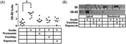
- 2 Insulin increases SR-BI levels in the apical surface of CaCo-2 cells and requires PI3K, AKT, and mTOR activities. A, CaCo-2 cells were incubated with inhibitors for PI3K (wortmannin), AKT (triciribine), or mTOR (rapamycin) for 1 h and then stimulated with 100 nM insulin plus the different inhibitors for additional 24 h, as indicated in Section 2. Whole cell lysates of CaCo-2 cells were analyzed by Western blot. B, CaCo-2 cells were incubated with triciribine or rapamycin and insulin as indicated in (A) and then their cell surface proteins were biotinylated as indicated in Section 2. Biotinylated cell lysates were precipitated with streptavidin and analyzed by Western blot with antibodies to IR and SR-BI as detailed in Section 2. IR, insulin receptor; mTOR, mammalian target of rapamycin; PI3K, phosphatidylinositol 3-kinase; SR-BI, scavenger receptor, class B, type I
