 Explore
 Validate
 Learn
Learn Western blot
 Immunocytochemistry
 Immunohistochemistry
Antibody data
- Antibody Data
- Antigen structure
- References [17]
- Comments [0]
- Validations
- Western blot [1]
- Immunocytochemistry [1]
Submit
Validation data
Reference
Comment
Report error
- Product number
- HPA003097 - Provider product page

- Provider
- Atlas Antibodies
- Proper citation
- Atlas Antibodies Cat#HPA003097, RRID:AB_1079473
- Product name
- Anti-NF2
- Antibody type
- Polyclonal
- Description
- Polyclonal Antibody against Human NF2, Gene description: neurofibromin 2 (merlin), Alternative Gene Names: merlin, Validated applications: WB, IHC, ICC, Uniprot ID: P35240, Storage: Store at +4°C for short term storage. Long time storage is recommended at -20°C.
- Reactivity
- Human, Mouse, Rat
- Host
- Rabbit
- Conjugate
- Unconjugated
- Isotype
- IgG
- Vial size
- 100 µl
- Concentration
- 0.4 mg/ml
- Storage
- Store at +4°C for short term storage. Long time storage is recommended at -20°C.
- Handling
- The antibody solution should be gently mixed before use.
Submitted references Spatial Landscape of Malignant Pleural and Peritoneal Mesothelioma Tumor Immune Microenvironments
Genomic and Transcriptomic Analyses of Malignant Pleural Mesothelioma (MPM) Samples Reveal Crucial Insights for Preclinical Testing
CNK2 promotes cancer cell motility by mediating ARF6 activation downstream of AXL signalling
A prognostic model for tumor recurrence and progression after meningioma surgery: preselection for further molecular work-up
Gatekeeping role of Nf2 /Merlin in vascular tip EC induction through suppression of VEGFR2 internalization
Comprehensive Analysis of VEGFR2 Expression in HPV-Positive and -Negative OPSCC Reveals Differing VEGFR2 Expression Patterns
Diverse Resistance Mechanisms to the Third-Generation ALK Inhibitor Lorlatinib in ALK-Rearranged Lung Cancer
Differential Expression of NF2 in Neuroepithelial Compartments Is Necessary for Mammalian Eye Development
Synergistic effect of Nutlin-3 combined with MG-132 on schwannoma cells through restoration of merlin and p53 tumour suppressors
Neurofibromatosis type 2 tumor suppressor protein is expressed in oligodendrocytes and regulates cell proliferation and process formation.
Differential NF2 Gene Status in Sporadic Vestibular Schwannomas and its Prognostic Impact on Tumour Growth Patterns
p53 performs an essential role in mediating the oncogenic stimulus triggered by loss of expression of neurofibromatosis type 2 during in vitro tumor progression
A critical role for NF2 and the Hippo pathway in branching morphogenesis
Nf2–Yap signaling controls the expansion of DRG progenitors and glia during DRG development
Homeostatic control of Hippo signaling activity revealed by an endogenous activating mutation in YAP
Tumor suppressor Nf2 limits expansion of the neural progenitor pool by inhibiting Yap/Taz transcriptional coactivators
The Merlin/NF2 Tumor Suppressor Functions through the YAP Oncoprotein to Regulate Tissue Homeostasis in Mammals
Ma X, Lembersky D, Kim E, Becich M, Testa J, Bruno T, Osmanbeyoglu H
Cancer Research Communications 2024;4(8):2133-2146
Cancer Research Communications 2024;4(8):2133-2146
Genomic and Transcriptomic Analyses of Malignant Pleural Mesothelioma (MPM) Samples Reveal Crucial Insights for Preclinical Testing
Laure A, Rigutto A, Kirschner M, Opitz L, Grob L, Opitz I, Felley-Bosco E, Hiltbrunner S, Curioni-Fontecedro A
Cancers 2023;15(10):2813
Cancers 2023;15(10):2813
CNK2 promotes cancer cell motility by mediating ARF6 activation downstream of AXL signalling
Serwe G, Kachaner D, Gagnon J, Plutoni C, Lajoie D, Duramé E, Sahmi M, Garrido D, Lefrançois M, Arseneault G, Saba-El-Leil M, Meloche S, Emery G, Therrien M
Nature Communications 2023;14(1)
Nature Communications 2023;14(1)
A prognostic model for tumor recurrence and progression after meningioma surgery: preselection for further molecular work-up
Padevit L, Vasella F, Friedman J, Mutschler V, Jenkins F, Held U, Rushing E, Wirsching H, Weller M, Regli L, Neidert M
Frontiers in Oncology 2023;13
Frontiers in Oncology 2023;13
Gatekeeping role of Nf2 /Merlin in vascular tip EC induction through suppression of VEGFR2 internalization
Bae J, Yang M, Jeong S, Kim J, Hong S, Kim J, Kim Y, Koh G
Science Advances 2022;8(23)
Science Advances 2022;8(23)
Comprehensive Analysis of VEGFR2 Expression in HPV-Positive and -Negative OPSCC Reveals Differing VEGFR2 Expression Patterns
Uzun S, Korkmaz Y, Wuerdemann N, Arolt C, Puladi B, Siefer O, Dönmez H, Hufbauer M, Akgül B, Klussmann J, Huebbers C
Cancers 2021;13(20):5221
Cancers 2021;13(20):5221
Diverse Resistance Mechanisms to the Third-Generation ALK Inhibitor Lorlatinib in ALK-Rearranged Lung Cancer
Recondo G, Mezquita L, Facchinetti F, Planchard D, Gazzah A, Bigot L, Rizvi A, Frias R, Thiery J, Scoazec J, Sourisseau T, Howarth K, Deas O, Samofalova D, Galissant J, Tesson P, Braye F, Naltet C, Lavaud P, Mahjoubi L, Abou Lovergne A, Vassal G, Bahleda R, Hollebecque A, Nicotra C, Ngo-Camus M, Michiels S, Lacroix L, Richon C, Auger N, De Baere T, Tselikas L, Solary E, Angevin E, Eggermont A, Andre F, Massard C, Olaussen K, Soria J, Besse B, Friboulet L
Clinical Cancer Research 2020;26(1):242-255
Clinical Cancer Research 2020;26(1):242-255
Differential Expression of NF2 in Neuroepithelial Compartments Is Necessary for Mammalian Eye Development
Moon K, Kim H, Lee D, Rao M, Levine E, Lim D, Kim J
Developmental Cell 2018;44(1):13-28.e3
Developmental Cell 2018;44(1):13-28.e3
Synergistic effect of Nutlin-3 combined with MG-132 on schwannoma cells through restoration of merlin and p53 tumour suppressors
Chen H, Xue L, Huang H, Wang H, Zhang X, Zhu W, Wang Z, Wang Z, Wu H
EBioMedicine 2018;36
EBioMedicine 2018;36
Neurofibromatosis type 2 tumor suppressor protein is expressed in oligodendrocytes and regulates cell proliferation and process formation.
Toledo A, Grieger E, Karram K, Morrison H, Baader SL
PloS one 2018;13(5):e0196726
PloS one 2018;13(5):e0196726
Differential NF2 Gene Status in Sporadic Vestibular Schwannomas and its Prognostic Impact on Tumour Growth Patterns
Chen H, Xue L, Wang H, Wang Z, Wu H
Scientific Reports 2017;7(1)
Scientific Reports 2017;7(1)
p53 performs an essential role in mediating the oncogenic stimulus triggered by loss of expression of neurofibromatosis type 2 during in vitro tumor progression
Li X, Chen H, Xue L, Pang X, Zhang X, Zhu Z, Zhu W, Wang Z, Wu H
Oncology Letters 2017;14(2):2223-2231
Oncology Letters 2017;14(2):2223-2231
A critical role for NF2 and the Hippo pathway in branching morphogenesis
Reginensi A, Enderle L, Gregorieff A, Johnson R, Wrana J, McNeill H
Nature Communications 2016;7(1)
Nature Communications 2016;7(1)
Nf2–Yap signaling controls the expansion of DRG progenitors and glia during DRG development
Serinagaoglu Y, Paré J, Giovannini M, Cao X
Developmental Biology 2015;398(1):97-109
Developmental Biology 2015;398(1):97-109
Homeostatic control of Hippo signaling activity revealed by an endogenous activating mutation in YAP
Chen Q, Zhang N, Xie R, Wang W, Cai J, Choi K, David K, Huang B, Yabuta N, Nojima H, Anders R, Pan D
Genes & Development 2015;29(12):1285-1297
Genes & Development 2015;29(12):1285-1297
Tumor suppressor Nf2 limits expansion of the neural progenitor pool by inhibiting Yap/Taz transcriptional coactivators
Lavado A, He Y, Paré J, Neale G, Olson E, Giovannini M, Cao X
Development 2013;140(16):3323-3334
Development 2013;140(16):3323-3334
The Merlin/NF2 Tumor Suppressor Functions through the YAP Oncoprotein to Regulate Tissue Homeostasis in Mammals
Zhang N, Bai H, David K, Dong J, Zheng Y, Cai J, Giovannini M, Liu P, Anders R, Pan D
Developmental Cell 2010;19(1):27-38
Developmental Cell 2010;19(1):27-38
No comments: Submit comment
Enhanced validation
- Submitted by
- Atlas Antibodies (provider)
- Enhanced method
- Genetic validation
- Main image
- Experimental details
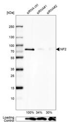
- Western blot analysis in U2OS cells transfected with control siRNA, target specific siRNA probe #1 and #2, using Anti-NF2 antibody. Remaining relative intensity is presented. Loading control: Anti-GAPDH.
- Sample type
- Human
- Protocol
- Protocol

Supportive validation
- Submitted by
- Atlas Antibodies (provider)
- Main image
- Experimental details
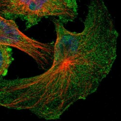
- Immunofluorescent staining of human cell line U-251 MG shows localization to plasma membrane & cytosol.
- Sample type
- Human
