Explore
 Validate
 Learn
Learn Western blot
 Immunohistochemistry
Antibody data
- Antibody Data
- Antigen structure
- References [2]
- Comments [0]
- Validations
- Western blot [1]
- Immunocytochemistry [1]
Submit
Validation data
Reference
Comment
Report error
- Product number
- GTX23546 - Provider product page

- Provider
- GeneTex
- Proper citation
- GeneTex Cat#GTX23546, RRID:AB_385098
- Product name
- beta 1 Adrenergic Receptor antibody
- Antibody type
- Polyclonal
- Reactivity
- Human, Mouse, Rat, Feline, Simian
- Host
- Rabbit
Submitted references Sympathetic innervation contributes to perineural invasion of salivary adenoid cystic carcinoma via the β2-adrenergic receptor.
Increased Efferent Cardiac Sympathetic Nerve Activity and Defective Intrinsic Heart Rate Regulation in Type 2 Diabetes.
Ma C, Gao T, Ju J, Zhang Y, Ni Q, Li Y, Zhao Z, Chai J, Yang X, Sun M
OncoTargets and therapy 2019;12:1475-1495
OncoTargets and therapy 2019;12:1475-1495
Increased Efferent Cardiac Sympathetic Nerve Activity and Defective Intrinsic Heart Rate Regulation in Type 2 Diabetes.
Thaung HP, Baldi JC, Wang HY, Hughes G, Cook RF, Bussey CT, Sheard PW, Bahn A, Jones PP, Schwenke DO, Lamberts RR
Diabetes 2015 Aug;64(8):2944-56
Diabetes 2015 Aug;64(8):2944-56
No comments: Submit comment
Supportive validation
- Submitted by
- GeneTex (provider)
- Main image
- Experimental details
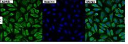
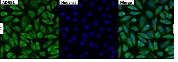
- Immunofluorescent analysis of ADR-Beta-1 (green) in untreated HeLa cells. Formalin fixed cells were permeabilized with 0.1% Triton X-100 in TBS for 15 minutes at room temperature. Cells were then blocked with 5% normal goat serum for 15 minutes at room temperature. Cells were probed with a rabbit polyclonal antibody recognizing ADR-Beta-1 (GTX23546), at a dilution of 1:100 for at least 1 hour at room temperature. Cells were washed with PBS and incubated with DyLight 488 goat-anti-rabbit secondary antibody at a dilution of 1:400 for 30 minutes at room temperature. Nuclei were stained with Hoechst 33342 dye.
Supportive validation
- Submitted by
- GeneTex (provider)
- Main image

- Experimental details
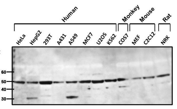
- Western blot analysis of ADR-Beta-1 was performed by loading 25ug of various whole cell lysates onto a 4-20% Tris-HCl polyacrylamide gel. Proteins were transferred to a PVDF membrane and blocked with 5% Milk/TBST for at least 1 hour. Membranes were probed with a rabbit polyclonal antibody recognizing ADR-Beta-1 (GTX23546) at a dilution of 1:1000 overnight at 4¢XC on a rocking platform.