Explore
 Validate
 Learn
Learn Western blot
 Immunoprecipitation
Antibody data
- Antibody Data
- Antigen structure
- References [1]
- Comments [0]
- Validations
- Western blot [1]
- Immunocytochemistry [1]
- Immunohistochemistry [2]
- Other assay [6]
Submit
Validation data
Reference
Comment
Report error
- Product number
- PA5-17034 - Provider product page

- Provider
- Invitrogen Antibodies
- Product name
- PIWIL1 Polyclonal Antibody
- Antibody type
- Polyclonal
- Antigen
- Synthetic peptide
- Description
- It is not recommended to aliquot this antibody.
- Reactivity
- Human, Mouse, Rat
- Host
- Rabbit
- Isotype
- IgG
- Vial size
- 100 µL
- Concentration
- 85 µg/mL
- Storage
- -20°C
Submitted references piRNA-associated proteins and retrotransposons are differentially expressed in murine testis and ovary of aryl hydrocarbon receptor deficient mice.
Rico-Leo EM, Moreno-Marín N, González-Rico FJ, Barrasa E, Ortega-Ferrusola C, Martín-Muñoz P, Sánchez-Guardado LO, Llano E, Alvarez-Barrientos A, Infante-Campos A, Catalina-Fernández I, Hidalgo-Sánchez M, de Rooij DG, Pendás AM, Peña FJ, Merino JM, Fernández-Salguero PM
Open biology 2016 Dec;6(12)
Open biology 2016 Dec;6(12)
No comments: Submit comment
Supportive validation
- Submitted by
- Invitrogen Antibodies (provider)
- Main image
- Experimental details
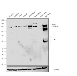
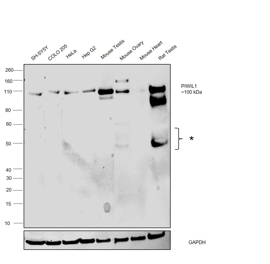
- Western Blot was performed using Anti-PIWIL1 Polyclonal Antibody (Product # PA5-17034) and a 100 kDa band corresponding to Piwi-like protein 1 was observed across the cell lines and tissues tested except Mouse Heart along with uncharacterized bands ~50 kDa. Whole cell extracts (30 µg lysate) of SH-SY5Y (Lane 1), COLO 205 (Lane 2), HeLa (Lane 3), Hep G2 (Lane 4) and tissue extracts of Mouse Testis (Lane 5), Mouse Ovary (Lane 6), Mouse Heart (Lane 7) and Rat Testis (Lane 8) were electrophoresed using NuPAGE™ 4-12% Bis-Tris Protein Gel (Product # NP0321BOX). Resolved proteins were then transferred onto a PVDF membrane (Product # LC2001) by iBlot® 2 Dry Blotting System (Product # IB21001). The blot was probed with the primary antibody (1:1000 dilution) and detected by chemiluminescence with Goat anti-Rabbit IgG (H+L) Superclonal™ Recombinant Secondary Antibody, HRP (Product # A27036, 1:4000 dilution) using the iBright FL 1000 (Product # A32752). Chemiluminescent detection was performed using SuperSignal™ West Atto Ultimate Sensitivity Substrate (Product # A38556).
Supportive validation
- Submitted by
- Invitrogen Antibodies (provider)
- Main image

- Experimental details
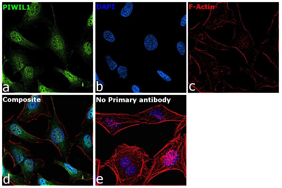
- Immunofluorescence analysis of Piwi-like protein 1 was performed using 70% confluent log phase HeLa cells. The cells were fixed with 4% paraformaldehyde for 15 minutes, permeabilized with 0.1% Triton™ X-100 for 15 minutes, and blocked with 2% BSA for 45 minutes at room temperature. The cells were labeled with PIWIL1 Polyclonal Antibody (Product # PA5-17034) at 1:200 dilution in 0.1% BSA, incubated at 4 degree celsius overnight and then labeled with Donkey anti-Rabbit IgG (H+L) Highly Cross-Adsorbed Secondary Antibody, Alexa Fluor Plus 488 (Product # A32790), (1:2000 dilution), for 45 minutes at room temperature (Panel a: Green). Nuclei (Panel b:Blue) were stained with ProLong™ Diamond Antifade Mountant with DAPI (Product # P36962). F-actin (Panel c: Red) was stained with Rhodamine Phalloidin (Product # R415, 1:300 dilution). Panel d represents the merged image showing nuclear and cytoplasmic localization. Panel e represents control HeLa cells with no primary antibody to assess background. The images were captured at 60X magnification.
Supportive validation
- Submitted by
- Invitrogen Antibodies (provider)
- Main image

- Experimental details
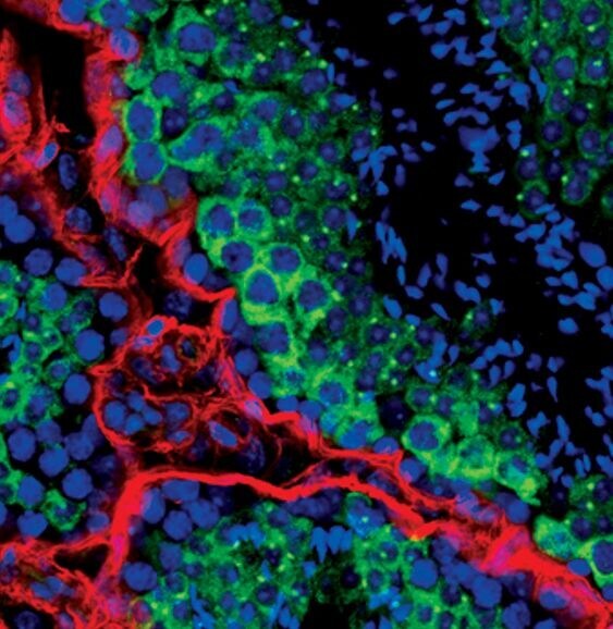
- Immunofluorescent analysis of Miwi in mouse testis using a Miwi polyclonal antibody (Product # PA5-17034) (green) and a Pan-Keratin monoclonal antibody (red). DNA is labeled using a fluorescent blue dye.
- Submitted by
- Invitrogen Antibodies (provider)
- Main image
- Experimental details
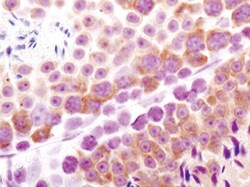
- Immunohistochemical analysis of Miwi in paraffin-embedded mouse testis using a Miwi polyclonal antibody (Product # PA5-17034).

Supportive validation
- Submitted by
- Invitrogen Antibodies (provider)
- Main image
- Experimental details
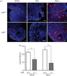
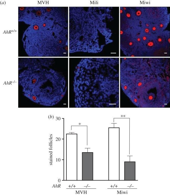
- Figure 9. Adult AhR- mice have reduced numbers of MVH- and Miwi-positive ovarian follicles. ( a ) Ovaries from adult (five to six weeks) AhR +/+ and AhR -/- mice were extracted and processed for the detection of MVH, Mili and Miwi by immunofluorescence. An Alexa-633-labelled secondary antibody has been used. ( b ) The number of positive follicles was quantified for each individual ovary of each mouse genotype. Ovaries from at least four AhR +/+ and AhR -/- mice were used, and immunofluorescences were done in triplicate. Data in panel ( b ) are shown as mean +- s.d. * p < 0.05, ** p < 0.01. Adult mice were 12-13 weeks of age. Scale bar, 50 um.
- Submitted by
- Invitrogen Antibodies (provider)
- Main image
- Experimental details
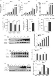
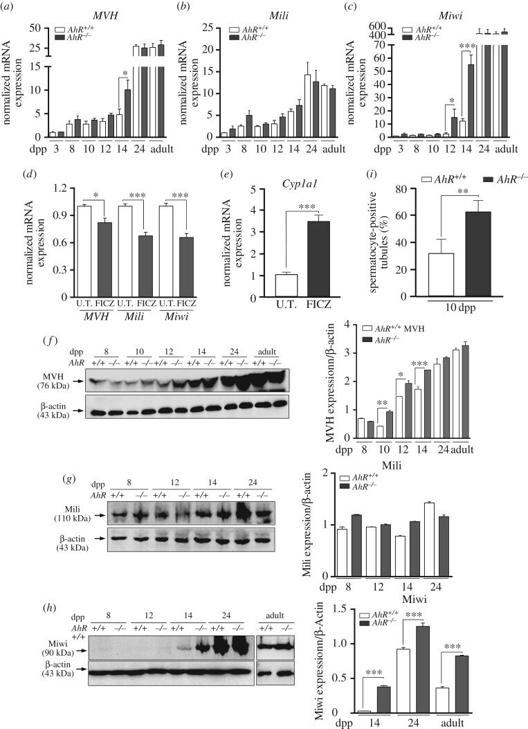
- Figure 2. AhR modulates the expression of piRNA-associated proteins MVH, Mili and Miwi. ( a - c ) Testes were obtained from AhR +/+ and AhR -/- mice at 3, 8, 10, 12, 14 and 24 dpp and from adults and used to isolate RNA. mRNA expression for MVH ( a ), Mili ( b ) and Miwi ( c ) was determined by RT-qPCR, using the oligonucleotides indicated in Material and methods. ( d ) AhR +/+ mice were subjected to intratesticular injection of 4 ug kg -1 of 6-formylindolo[3,2-b]carbazole (FICZ) to activate AhR in vivo and the mRNA expression of Mvh , Mili and Miwi analysed by RT-qPCR. ( e ) The efficiency of FICZ to induce AhR in the testis was analysed by measuring the mRNA expression of its canonical target gene Cyp1a1 . Gene expression in panels ( a-e ) has been normalized by Gapdh and represented as 2 -DeltaDeltaCt . ( f-h ) Testes from AhR +/+ and AhR -/- mice were obtained and total protein extracts prepared and analysed for the expression of MVH ( f ), Mili ( g ) and Miwi ( h ) by immunoblotting using specific antibodies. beta-Actin was used to normalize protein levels. ( i ) The maturation of AhR +/+ and AhR -/- testes at 10 dpp was determined by the microscopic quantification of spermatocyte-positive seminiferous tubules in at least six different male mice of each genotype. At least four individual mice of each genotype were used for the experiments shown in panels ( a-h ). Determinations were done in duplicate. Data are shown as mean +- s.d. * p
- Submitted by
- Invitrogen Antibodies (provider)
- Main image
- Experimental details
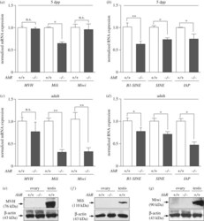
- Figure 8. AhR deficiency reduces the levels of transposons and piRNA-associated proteins in the ovary. ( a ) Total RNA was isolated from ovaries of 5 dpp AhR +/+ and AhR -/- mice and used to analyse by RT-qPCR the mRNA expression of piRNA-associated proteins MVH , Mili and Miwi . ( b ) Total RNAs were also used to quantify by RT-qPCR the levels of retrotransposons of the SINE and IAP families and B1-SINE subfamily . ( c ) Ovaries from adult (five to six weeks) AhR +/+ and AhR -/- mice were used to isolate total RNA. The mRNA levels of MVH, Mili and Miwi were determined by RT-qPCR. ( d ) Total RNAs were also used to analyse by RT-qPCR the expression of B1-SINE , SINE and IAP retrotransposons in adult AhR +/+ and AhR -/- ovaries. Gene expression has been normalized by Gapdh and represented as 2 -DeltaDeltaCt . ( e-g ) Protein expression of MVH, Mili and Miwi was analysed by immunoblotting using total ovary protein extracts from adult AhR +/+ and AhR -/- mice. Experiments were done in duplicate in four biological replicates. The expression of beta-actin was used to normalize protein loading. Data in panels ( a-d ) are shown as mean +- s.d. * p < .05, ** p < 0.01. n.s . , not statistically significant. Adult mice were 12-13 weeks of age.

- Submitted by
- Invitrogen Antibodies (provider)
- Main image
- Experimental details
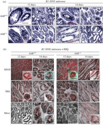
- Figure 5. B1-SINE retrotransposons and piRNA-associated proteins have similar expression patterns in mouse testes. ( a ) AhR +/+ and AhR -/- testes obtained at 12 and 14 dpp were processed for in situ hybridization as indicated in Material and methods. Tissue sections were analysed for B1-SINE expression using the antisense sequence. ( b ) In situ hybridization for the B1-SINE retrotransposon was combined with immunofluorescence for MVH, Mili and Miwi in testis sections from AhR +/+ and AhR -/- mice at 12 and 14 dpp. Details of the micrographs are shown in the insets. In situ hybridization is shown in black in panel ( b ) to emphasize MVH, Miwi and Mili protein expression. Three biological replicates were used for each experiment. Scale bar, 50 um.

- Submitted by
- Invitrogen Antibodies (provider)
- Main image
- Experimental details
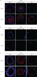
- Figure 10. Lack of AhR alters the pattern of MVH expression during oocyte maturation. ( a-c ) Oocytes at the follicle, GV and MII stages were extracted from AhR +/+ and AhR -/- mice as indicated in Material and methods. The protein expression of MVH ( a ), Mili ( b ) and Miwi ( c ) was analysed by immunofluorescence using specific antibodies. An Alexa-633-labelled secondary antibody has been used. Groups of 10 mice of each genotype were used to extract oocytes at the different developmental stages. Adult mice were 12-13 weeks of age. Scale bar, 50 um.

- Submitted by
- Invitrogen Antibodies (provider)
- Main image

- Experimental details
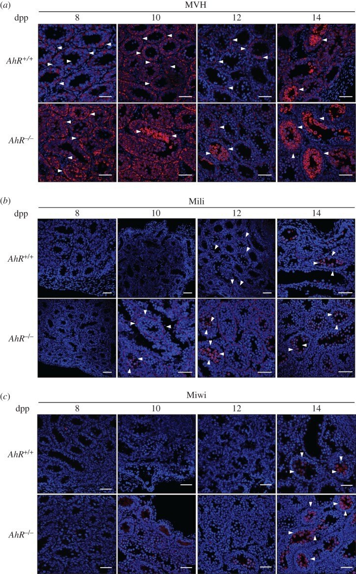
- Figure 3. AhR deficiency may accelerate the temporal expression pattern of MVH, Mili and Miwi. AhR +/+ and AhR -/- testes were extracted at 8, 10, 12 and 14 dpp and processed for immunohistochemistry as indicated in Material and methods. ( a ) Testis sections were stained for MVH using a specific antibody. ( b ) Sections were also analysed for the location of the Mili protein. ( c ) The pattern of Miwi expression was also determined in testis sections of both genotypes. DAPI staining was used to label cell nuclei. Arrowheads indicate protein expression. At least five individual mice of each genotype were used for the experiments, and different sections from each testis were analysed. Scale bar, 50 um.