Explore
 Validate
 Learn
Learn Western blot
Antibody data
- Antibody Data
- Antigen structure
- References [1]
- Comments [0]
- Validations
- Western blot [4]
- Immunohistochemistry [9]
Submit
Validation data
Reference
Comment
Report error
- Product number
- NBP1-90692 - Provider product page

- Provider
- Novus Biologicals
- Proper citation
- Novus Cat#NBP1-90692, RRID:AB_11020096
- Product name
- Rabbit Polyclonal UFL1 Antibody
- Antibody type
- Polyclonal
- Description
- Immunogen affinity purified. Specificity of human UFL1 antibody verified on a Protein Array containing target protein plus 383 other non-specific proteins.
- Reactivity
- Human
- Host
- Rabbit
- Isotype
- IgG
- Vial size
- 0.1 ml
- Storage
- Store at 4C short term. Aliquot and store at -20C long term. Avoid freeze-thaw cycles.
Submitted references Functional CRISPR screening identifies the ufmylation pathway as a regulator of SQSTM1/p62.
DeJesus R, Moretti F, McAllister G, Wang Z, Bergman P, Liu S, Frias E, Alford J, Reece-Hoyes JS, Lindeman A, Kelliher J, Russ C, Knehr J, Carbone W, Beibel M, Roma G, Ng A, Tallarico JA, Porter JA, Xavier RJ, Mickanin C, Murphy LO, Hoffman GR, Nyfeler B
eLife 2016 Jun 28;5
eLife 2016 Jun 28;5
No comments: Submit comment
Supportive validation
- Submitted by
- Novus Biologicals (provider)
- Main image

- Experimental details
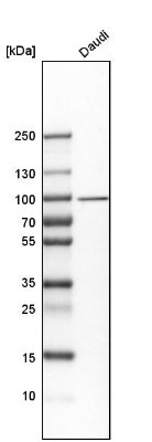
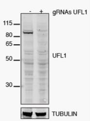
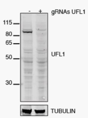
- Western Blot: UFL1 Antibody [NBP1-90692] - Analysis in human cell line Daudi.
- Submitted by
- Novus Biologicals (provider)
- Main image
- Experimental details
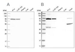
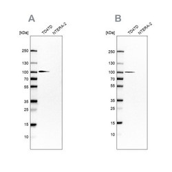
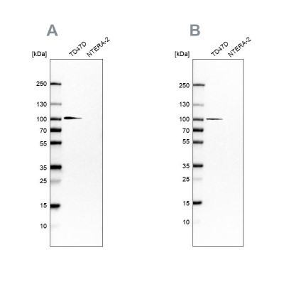
- Western Blot: UFL1 Antibody [NBP1-90692] - Analysis using Anti-UFL1 antibody NBP1-90692 (A) shows similar pattern to independent antibody NBP1-90691 (B).
- Submitted by
- Novus Biologicals (provider)
- Main image
- Experimental details
- Western Blot: UFL1 Antibody [NBP1-90692] - HeLa cell lysate (35 ug each lane). Image from verified customer review.
- Submitted by
- Novus Biologicals (provider)
- Main image
- Experimental details
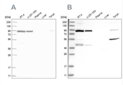
- Western Blot: UFL1 Antibody [NBP1-90692] - Analysis using Anti-UFL1 antibody NBP1-90692 (A) shows similar pattern to independent antibody NBP1-90691 (B).
Supportive validation
- Submitted by
- Novus Biologicals (provider)
- Main image
- Experimental details
- Immunohistochemistry-Paraffin: UFL1 Antibody [NBP1-90692] - Staining of human colon using Anti-UFL1 antibody HPA030560.
- Submitted by
- Novus Biologicals (provider)
- Main image

- Experimental details
- Immunohistochemistry-Paraffin: UFL1 Antibody [NBP1-90692] - Staining of human kidney using Anti-UFL1 antibody.
- Submitted by
- Novus Biologicals (provider)
- Main image
- Experimental details
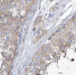
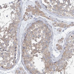
- Immunohistochemistry-Paraffin: UFL1 Antibody [NBP1-90692] - Staining of human prostate using Anti-UFL1 antibody.
- Submitted by
- Novus Biologicals (provider)
- Main image
- Experimental details
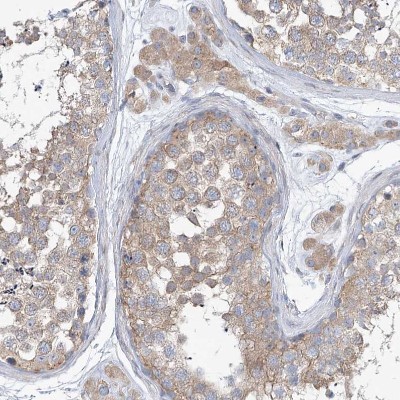
- Immunohistochemistry-Paraffin: UFL1 Antibody [NBP1-90692] - Staining of human testis using Anti-UFL1 antibody.

- Submitted by
- Novus Biologicals (provider)
- Main image

- Experimental details
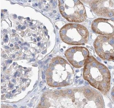
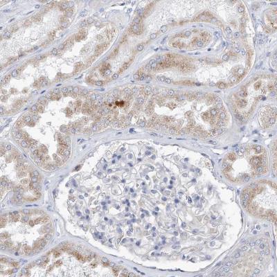
- Immunohistochemistry: UFL1 Antibody [NBP1-90692] - Immunohistochemical staining of human kidney shows very weak cytoplasmic positivity in cells in tubules.
- Submitted by
- Novus Biologicals (provider)
- Main image
- Experimental details
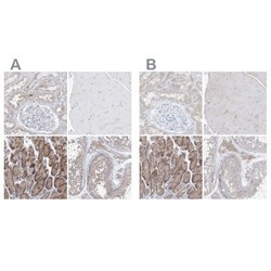
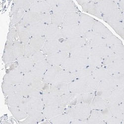
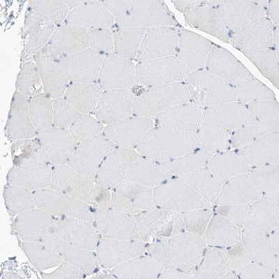
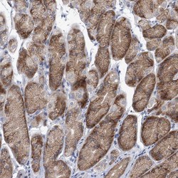
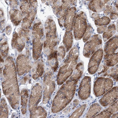
- Immunohistochemistry-Paraffin: UFL1 Antibody [NBP1-90692] - Staining of human kidney, skeletal muscle, stomach and testis using Anti-UFL1 antibody NBP1-90692 (A) shows similar protein distribution across tissues to independent antibody NBP1-90691 (B).

- Submitted by
- Novus Biologicals (provider)
- Main image
- Experimental details
- Immunohistochemistry-Paraffin: UFL1 Antibody [NBP1-90692] - Staining of human skeletal muscle shows no positivity in myocytes as expected.
- Submitted by
- Novus Biologicals (provider)
- Main image
- Experimental details
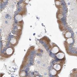
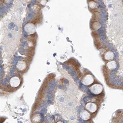
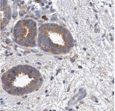
- Immunohistochemistry-Paraffin: UFL1 Antibody [NBP1-90692] - Staining of human stomach shows weak to moderate cytoplasmic positivity in glandular cells.
- Submitted by
- Novus Biologicals (provider)
- Main image
- Experimental details
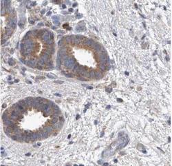
- Immunohistochemistry-Paraffin: UFL1 Antibody [NBP1-90692] - Staining of human testis shows weak to moderate cytoplasmic positivity in Leydig cells and cells in seminifereus ducts.