Explore
 Validate
 Learn
Learn Flow cytometry
Antibody data
- Antibody Data
- Antigen structure
- References [2]
- Comments [0]
- Validations
- Flow cytometry [1]
- Other assay [4]
Submit
Validation data
Reference
Comment
Report error
- Product number
- 51-8828-41 - Provider product page

- Provider
- Invitrogen Antibodies
- Product name
- Granulysin Monoclonal Antibody (eBioDH2 (DH2)), Alexa Fluor™ 647, eBioscience™
- Antibody type
- Monoclonal
- Antigen
- Other
- Description
- Description: The eBioDH2 monoclonal antibody reacts with human granulysin, a protein involved in the cytolytic pathway. Granulysin is a member of the saposin-like protein (SAPLIP) family. Two proteins 9 and 15kDa are thought to be post translation modified versions of the 15kDa protein with a change in confirmation. Granulysin is found in NK and activated T cells and the expression is confined to the cytolytic granules which also contains perforin and granzyme. Granulysin with perforn are suspected to function synergistically to kill bacteria and induce apoptosis in nucleated cells.
- Conjugate
- Red dye
- Antibody clone number
- eBioDH2 (DH2)
- Concentration
- 5 µL/Test
Submitted references Nanocages engineered from Bacillus Calmette-Guerin facilitate protective Vγ2Vδ2 T cell immunity against Mycobacterium tuberculosis infection.
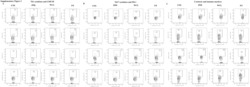
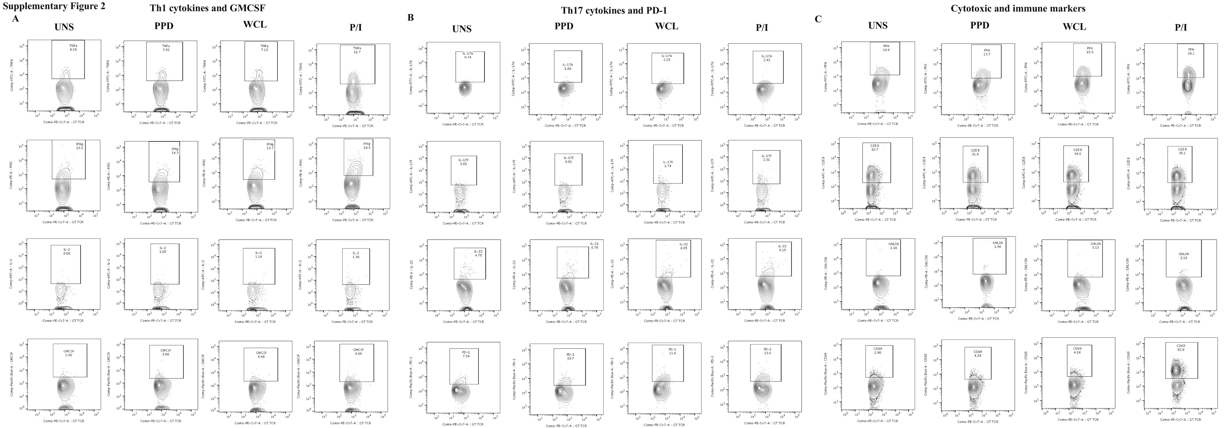
Decreased Frequencies of Gamma/Delta T Cells Expressing Th1/Th17 Cytokine, Cytotoxic, and Immune Markers in Latent Tuberculosis-Diabetes/Pre-Diabetes Comorbidity.
Pi J, Zhang Z, Yang E, Chen L, Zeng L, Chen Y, Wang R, Huang D, Fan S, Lin W, Shen H, Xu JF, Zeng G, Shen L
Journal of nanobiotechnology 2022 Jan 15;20(1):36
Journal of nanobiotechnology 2022 Jan 15;20(1):36
Decreased Frequencies of Gamma/Delta T Cells Expressing Th1/Th17 Cytokine, Cytotoxic, and Immune Markers in Latent Tuberculosis-Diabetes/Pre-Diabetes Comorbidity.
Kathamuthu GR, Kumar NP, Moideen K, Menon PA, Babu S
Frontiers in cellular and infection microbiology 2021;11:756854
Frontiers in cellular and infection microbiology 2021;11:756854
No comments: Submit comment
Supportive validation
- Submitted by
- Invitrogen Antibodies (provider)
- Main image
- Experimental details
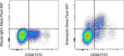
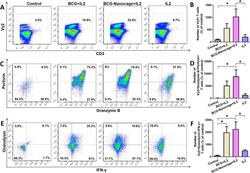
- Normal human peripheral blood cells were stained intracellularly, using the Intracellular Fixation & Permeabilization Buffer Set (Product # 88-8824-00) and protocol, with CD56 Monoclonal Antibody, FITC (Product # 11-0566-42) and Mouse IgG1 kappa Isotype Control, Alexa Fluor 647 (Product # 51-4714-81) (left) or Granulysin Monoclonal Antibody, Alexa Fluor 647 (right). Cells in the lymphocyte gate were used for analysis.
- Conjugate
- Red dye

Supportive validation
- Submitted by
- Invitrogen Antibodies (provider)
- Main image
- Experimental details
- NULL
- Conjugate
- Red dye
- Submitted by
- Invitrogen Antibodies (provider)
- Main image
- Experimental details
- NULL
- Conjugate
- Red dye
- Submitted by
- Invitrogen Antibodies (provider)
- Main image

- Experimental details
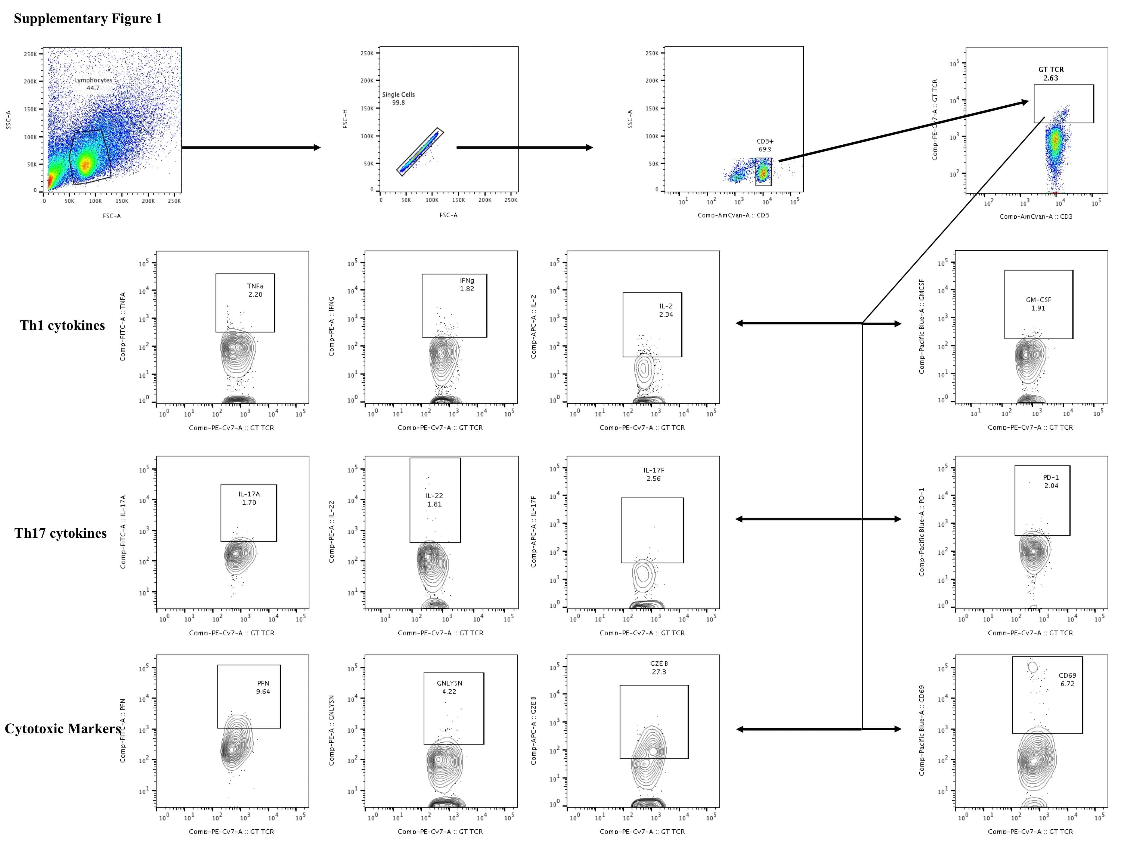
- Figure 3 Decreased frequencies of gammadelta T cells expressing cytotoxic markers in LTB comorbidities. PBMCs were either untreated or treated with Mtb or positive control antigens for 18 h. The absolute (unstimulated, UNS) and antigen-stimulated (PPD, WCL, P/I) net frequencies of cytotoxic (PFN, GZEB, GNLYSN) markers were shown in LTB DM (n = 20), LTB PDM (n = 20), and LTB NDM (n = 20) groups. Geometric mean values were represented using bars, and every circle denotes a single individual. Kruskal-Wallis test with multiple Dunn's comparison were used to determine the p values.
- Conjugate
- Red dye
- Submitted by
- Invitrogen Antibodies (provider)
- Main image
- Experimental details
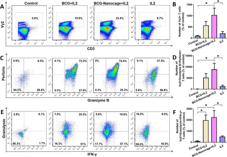
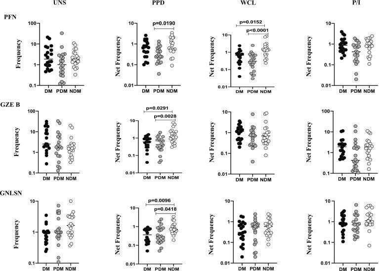
- Fig. 3 Effect BCG-Nanocage on e x vivo activation and expansion of Vgamma2+ T cells in PBMC from non-human primates. A Typical flow cytometry panel for Vgamma2+ cells gated in T cells from PBMC with or without (co-)stimulation using BCG/IL2, BCG-Nanocage/IL2 or IL2 for 6 days. B Effects of (co-)stimulation using BCG/IL2, BCG-Nanocage/IL2 or IL2 on Vgamma2+ T cells after 6 day treatment, n = 3, *p < 0.05. C Typical flow cytometry panel for Granzyme B+ Perforin+ cells gated in Vgamma2+ T cells in total T cells from PBMC with or without (co-)stimulation using BCG/IL2, BCG-Nanocage/IL2 or IL2 for 6 days. D Effects of (co-)stimulation using BCG/IL2, BCG-Nanocage/IL2 or IL2 on Vgamma2+ Granzyme B+ Perforin+ cells in T cells after 6 day treatment, n = 3, *p < 0.05. E Typical flow cytometry panel for Granulysin + IFN-gamma + cells gated in Vgamma2+ T cells from PBMC with or without (co-)stimulation using BCG/IL2, BCG-Nanocage/IL2 or IL2 for 6 days. F Effects of (co-)stimulation using BCG/IL2, BCG-Nanocage/IL2 or IL2 on Vgamma2+ Granulysin + IFN-gamma+ cells in T cells after 6 day treatment, n = 3, *p < 0.05
- Conjugate
- Red dye