Explore
 Validate
 Learn
Learn Immunocytochemistry
Antibody data
- Antibody Data
- Antigen structure
- References [1]
- Comments [0]
- Validations
- Immunocytochemistry [1]
- Other assay [1]
Submit
Validation data
Reference
Comment
Report error
- Product number
- PA5-36003 - Provider product page

- Provider
- Invitrogen Antibodies
- Product name
- TRUB1 Polyclonal Antibody
- Antibody type
- Polyclonal
- Antigen
- Recombinant full-length protein
- Description
- Store product as a concentrated solution. Centrifuge briefly prior to opening the vial. Predicted reactivity: Mouse (91%), Rat (92%), Rhesus Monkey (98%).
- Reactivity
- Human
- Host
- Rabbit
- Isotype
- IgG
- Vial size
- 100 μL
- Concentration
- 1 mg/mL
- Storage
- Store at 4°C short term. For long term storage, store at -20°C, avoiding freeze/thaw cycles.
Submitted references Human TRUB1 is a highly conserved pseudouridine synthase responsible for the formation of Ψ55 in mitochondrial tRNAAsn, tRNAGln, tRNAGlu and tRNAPro.
Jia Z, Meng F, Chen H, Zhu G, Li X, He Y, Zhang L, He X, Zhan H, Chen M, Ji Y, Wang M, Guan MX
Nucleic acids research 2022 Sep 9;50(16):9368-9381
Nucleic acids research 2022 Sep 9;50(16):9368-9381
No comments: Submit comment
Supportive validation
- Submitted by
- Invitrogen Antibodies (provider)
- Main image
- Experimental details
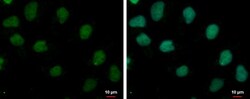
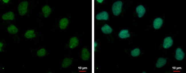
- TRUB1 Polyclonal Antibody detects TRUB1 protein at nucleus by immunofluorescent analysis. Sample: NT2D1 cells were fixed in 4% paraformaldehyde at RT for 15 min. Green: TRUB1 protein stained by TRUB1 Polyclonal Antibody (Product # PA5-36003) diluted at 1:500. Blue: Hoechst 33342 staining.
Supportive validation
- Submitted by
- Invitrogen Antibodies (provider)
- Main image
- Experimental details
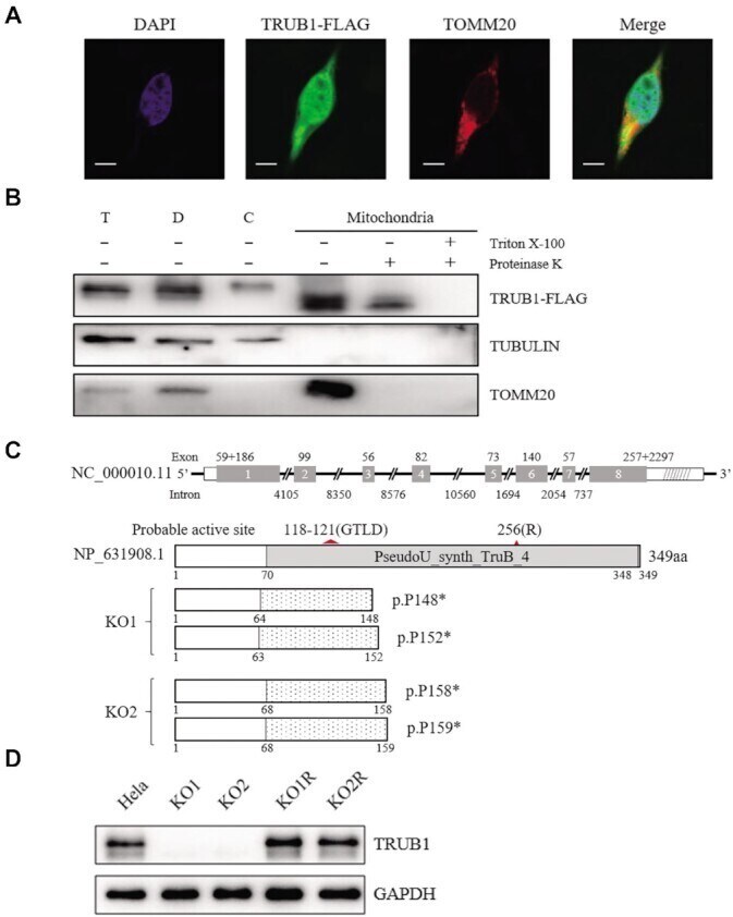
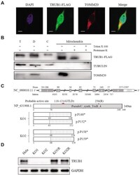
- Figure 2. Subcellular location and generation of TRUB1 knockout HeLa cell lines using CRISPR/Cas9 system. ( A ) Subcellular localization of TRUB1 by immunofluorescence in HeLa cells. TRUB1-FLAG (shown in green), TOMM20 (shown in red), and DAPI (shown in blue). Scale bar: 10 mum. ( B ) Subcellular localization of TRUB1 by Western blot with anti-FLAG, TOMM20 (mitochondrial) and TUBULIN (cytosol). T, total cell lysate; D, debris; C, cytosol; Mito, mitochondria. Isolated mitochondria were treated with (+) or without (-) 1% Triton X-100 followed by proteinase K digestion, respectively. ( C ) Schematic representation of TRUB1 and its truncated proteins. Shaded boxes indicate the PseudoU_synth_TruB_4 domain of TRUB1. Red triangle shows the probable active site. Deletion or insertion resulting in truncated proteins. ( D ) Western blot analysis. Twenty micrograms of total cellular proteins of each cell line were electrophoresed through and hybridized with antibodies specific for TRUB1 or with GAPDH as a loading control. KO1 and KO2 represented two TRUB1 knockout - cell lines; KO1R and KO2R represented KO1 and KO2 expressing wild type TRUB1 cDNA.