 Explore
 Validate
 Learn
Learn Western blot
Antibody data
- Antibody Data
- Antigen structure
- References [4]
- Comments [0]
- Validations
- Western blot [1]
- Immunohistochemistry [1]
Submit
Validation data
Reference
Comment
Report error
- Product number
- AF5810 - Provider product page

- Provider
- R&D Systems
- Product name
- Human/Mouse WDR5 Antibody
- Antibody type
- Polyclonal
- Description
- Antigen Affinity-purified. Detects human and mouse WDR5 in Western blots.
- Reactivity
- Human, Mouse
- Host
- Goat
- Conjugate
- Unconjugated
- Antigen sequence
P61964- Isotype
- IgG
- Vial size
- 100 ug
- Concentration
- LYOPH
- Storage
- Use a manual defrost freezer and avoid repeated freeze-thaw cycles. 12 months from date of receipt, -20 to -70 °C as supplied. 1 month, 2 to 8 °C under sterile conditions after reconstitution. 6 months, -20 to -70 °C under sterile conditions after reconstitution.
Submitted references Expression of WD Repeat Domain 5 (WDR5) is Associated with Progression and Reduced Prognosis in Papillary Thyroid Carcinoma.
PI3K/AKT-mediated upregulation of WDR5 promotes colorectal cancer metastasis by directly targeting ZNF407.
GRHL3/GET1 and trithorax group members collaborate to activate the epidermal progenitor differentiation program.
Essential role of the CUL4B ubiquitin ligase in extra-embryonic tissue development during mouse embryogenesis.
Xu W, Wang L, An Y, Ye J
Medical science monitor : international medical journal of experimental and clinical research 2019 May 20;25:3762-3770
Medical science monitor : international medical journal of experimental and clinical research 2019 May 20;25:3762-3770
PI3K/AKT-mediated upregulation of WDR5 promotes colorectal cancer metastasis by directly targeting ZNF407.
Tan X, Chen S, Wu J, Lin J, Pan C, Ying X, Pan Z, Qiu L, Liu R, Geng R, Huang W
Cell death & disease 2017 Mar 16;8(3):e2686
Cell death & disease 2017 Mar 16;8(3):e2686
GRHL3/GET1 and trithorax group members collaborate to activate the epidermal progenitor differentiation program.
Hopkin AS, Gordon W, Klein RH, Espitia F, Daily K, Zeller M, Baldi P, Andersen B
PLoS genetics 2012;8(7):e1002829
PLoS genetics 2012;8(7):e1002829
Essential role of the CUL4B ubiquitin ligase in extra-embryonic tissue development during mouse embryogenesis.
Liu L, Yin Y, Li Y, Prevedel L, Lacy EH, Ma L, Zhou P
Cell research 2012 Aug;22(8):1258-69
Cell research 2012 Aug;22(8):1258-69
No comments: Submit comment
Supportive validation
- Submitted by
- R&D Systems (provider)
- Main image

- Experimental details
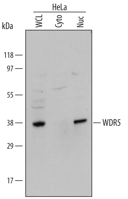
- Detection of Human WDR5 by Western Blot. Western blot shows lysates of HeLa human cervical epithelial carcinoma cell line. Gels were loaded with 30 μg of whole cell lysate (WCL), 20 μg of cytoplasmic (Cyto), and 10 μg of nuclear extracts (Nuc). PVDF membrane was probed with 1 µg/mL Goat Anti-Human/Mouse WDR5 Antigen Affinity-purified Polyclonal Antibody (Catalog # AF5810) followed by HRP-conjugated Anti-Goat IgG Secondary Antibody (Catalog # HAF017). A specific band for WDR5 was detected at approximately 40 kDa (as indicated). This experiment was conducted under reducing conditions and using Immunoblot Buffer Group 1.
Supportive validation
- Submitted by
- R&D Systems (provider)
- Main image
- Experimental details
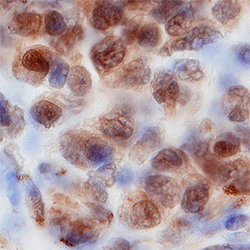
- WDR5 in Human B-Cell Lymphoma. WDR5 was detected in immersion fixed paraffin-embedded sections of human B-cell lymphoma using Goat Anti-Human/Mouse WDR5 Antigen Affinity-purified Polyclonal Antibody (Catalog # AF5810) at 15 µg/mL for 1 hour at room temperature followed by incubation with the Anti-Goat IgG VisUCyte™ HRP Polymer Antibody (Catalog # VC004). Tissue was stained using DAB (brown) and counterstained with hematoxylin (blue). Specific staining was localized to nuclei. View our protocol for IHC Staining with VisUCyte HRP Polymer Detection Reagents.
