 Explore
 Validate
 Learn
Learn Western blot
Antibody data
- Antibody Data
- Antigen structure
- References [1]
- Comments [0]
- Validations
- Western blot [6]
- Immunocytochemistry [3]
- Immunohistochemistry [1]
- Flow cytometry [1]
Submit
Validation data
Reference
Comment
Report error
- Product number
- 45-6400 - Provider product page

- Provider
- Invitrogen Antibodies
- Product name
- Mitofilin Monoclonal Antibody (2E4AD5)
- Antibody type
- Monoclonal
- Antigen
- Other
- Description
- Positive controls: Human fibroblasts; Human cerebellum tissue; Isolated mitochondria from Human, Bovine, Rat and Mouse hearts, HepG2 lysate; HL60 cells. This clone can also be referred to as clone number MM#7A-2E4AD5.
- Reactivity
- Human, Mouse, Rat, Bovine
- Host
- Mouse
- Isotype
- IgG
- Antibody clone number
- 2E4AD5
- Vial size
- 100 µg
- Concentration
- 1 mg/mL
- Storage
- 4° C, do not freeze
Submitted references Cardiac ischemia/reperfusion stress reduces inner mitochondrial membrane protein (mitofilin) levels during early reperfusion.
Tombo N, Imam Aliagan AD, Feng Y, Singh H, Bopassa JC
Free radical biology & medicine 2020 Oct;158:181-194
Free radical biology & medicine 2020 Oct;158:181-194
No comments: Submit comment
Supportive validation
- Submitted by
- Invitrogen Antibodies (provider)
- Main image
- Experimental details
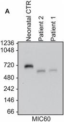
- Western blot analysis of Mitofilin in mitochondria using a Mitofilin Monoclonal antibody (Product # 45-6400). Lane 1: Control neonatal mitochondria from skin fibroblasts, Lane 2: Patient 2 (male) mitochondria from skin fibroblasts, Lane 3: Patient 1 (female) mitochondria from skin fibroblasts. Predicted band size: 84 kDa. Clinical findings for patients 1 & 2 suggest neonatal onset mitochondrial encephalopathy with recurrent bouts of liver disease.

- Submitted by
- Invitrogen Antibodies (provider)
- Main image

- Experimental details
- Knockdown of Mitofilin was achieved by transfecting HeLa with Mitofilin specific siRNAs (Silencer® select Product # S21634, S21635). Western blot analysis (Fig. a) was performed using Whole cell extracts from the Mitofilin knockdown cells (lane 3), non-targeting scrambled siRNA transfected cells (lane 2) and untransfected cells (lane 1). The blot was probed with Mitofilin Monoclonal Antibody (2E4AD5) (Product # 45-6400, 2 µg/mL dilution) and Goat anti-Mouse IgG (H+L) Superclonal™ Recombinant Secondary Antibody, HRP (Product # A28177, 1:10,000 dilution). Densitometric analysis of this western blot is shown in histogram (Fig. b). Decrease in signal upon siRNA mediated knock down confirms that antibody is specific to Mitofilin.

- Submitted by
- Invitrogen Antibodies (provider)
- Main image

- Experimental details
- Western blot was performed using Anti-Mitofilin Monoclonal Antibody (2E4AD5) (Product # 45-6400) and a 83 kDa band corresponding to Mitofilin was observed across all tested cell line lysates. Whole cell extracts (30 µg lysate) of MCF7 (Lane 1), A549 (Lane 2), HEK-293 (Lane 3), U-2 OS (Lane 4) and HeLa (Lane 5) were electrophoresed using NuPAGE™ 10% Bis-Tris Protein Gel (Product # NP0302BOX), 12 well. Resolved proteins were then transferred onto a nitrocellulose membrane (Product # IB23001) by iBlot® 2 Dry Blotting System (Product # IB21001). The blot was probed with the primary antibody (2 µg/mL dilution) and detected by chemiluminescence with Goat anti-Mouse IgG (H+L) Superclonal™ Recombinant Secondary Antibody, HRP (Product # A28177, 1:10,000 dilution) using the iBright™ FL1500 Imaging System (Product # A44115). Chemiluminescent detection was performed using SuperSignal™ West Pico PLUS Chemiluminescent Substrate (Product # 34580).

- Submitted by
- Invitrogen Antibodies (provider)
- Main image

- Experimental details
- Western blot analysis of Mitofilin in isolated mitochondria using a Mitofilin Monoclonal antibody (Product # 45-6400) at a concentration of 2 µg/mL. Lane 1: Isolated mitochondria from Human heart at 5 µg, Lane 2: Isolated mitochondria from Bovine heart at 1 µg, Lane 3: Isolated mitochondria from Rat heart at 10 µg, Lane 4: Isolated mitochondria from Mouse heart at 10 µg, Lane 5: HepG2 cell lysate at 20 µg. Predicted band size: 84 kDa. Extra bands in the mouse sample (lane 4) are due to the reaction of the IgG-specific goat anti-mouse secondary antibody with residual mouse blood in the heart tissue, as it is very difficult to entirely remove the blood from these small organs.

Supportive validation
- Submitted by
- Invitrogen Antibodies (provider)
- Main image

- Experimental details
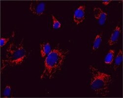
- Immunocytochemical analysis of Mitofilin in Cultured Human fibroblasts using an Mitofilin Monoclonal antibody (Product # 45-6400) at a concentration of 5 µg/mL. The Cultured Human fibroblasts were fixed, permeabilized, and a fluorescent goat-anti-mouse IgG secondary antibody was used.

- Submitted by
- Invitrogen Antibodies (provider)
- Main image

- Experimental details
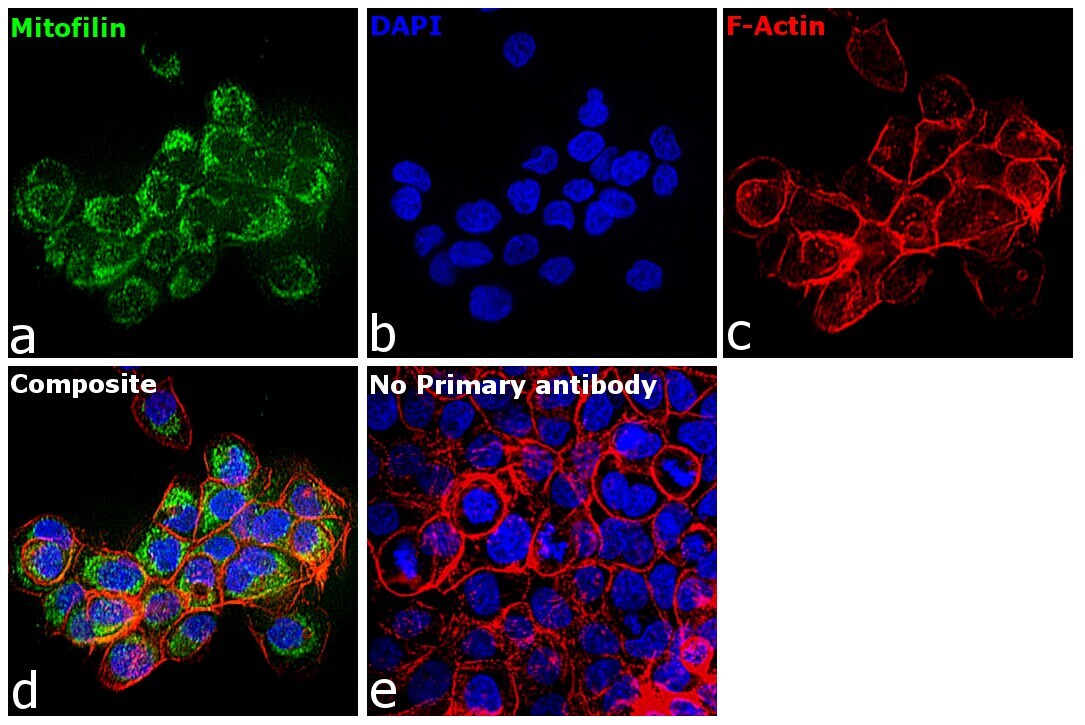
- Immunofluorescence analysis of Mitofilin was performed using 70% confluent log phase A-431 cells. The cells were fixed with 4% paraformaldehyde for 10 minutes, permeabilized with 0.1% Triton™ X-100 for 15 minutes, and blocked with 2% BSA for 1 hour at room temperature. The cells were labeled with Mitofilin Monoclonal Antibody (2E4AD5) (Product # 45-6400) at 1:100 dilution in 0.1% BSA, incubated at 4 degree celsius overnight and then labeled with Donkey anti-Mouse IgG (H+L) Highly Cross-Adsorbed Secondary Antibody, Alexa Fluor Plus 488 (Product # A32766, 1:2000), for 45 minutes at room temperature (Panel a: Green). Nuclei (Panel b:Blue) were stained with Hoechst 33342 (Product # H1399). F-actin (Panel c: Red) was stained with Alexa Fluor™ Plus 647 Phalloidin (Product # A30107, 1:2000 dilution). Panel d represents the merged image showing cytoplasmic (mitochondrial like) localization. Panel e represents control cells with no primary antibody to assess background. The images were captured at 40X magnification in CellInsight CX7 LZR High-Content Screening (HCS) Platform (Product # CX7A1110LZR) and externally deconvoluted (D.Sage et al. / Methods 115 (2017) 28–41).
- Submitted by
- Invitrogen Antibodies (provider)
- Main image
- Experimental details
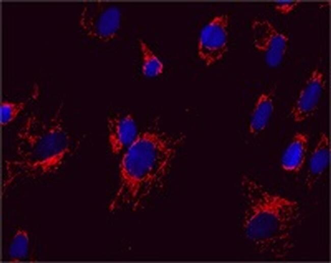
- Immunocytochemical analysis of Mitofilin in Cultured Human fibroblasts using an Mitofilin Monoclonal antibody (Product # 45-6400) at a concentration of 5 µg/mL. The Cultured Human fibroblasts were fixed, permeabilized, and a fluorescent goat-anti-mouse IgG secondary antibody was used.
Supportive validation
- Submitted by
- Invitrogen Antibodies (provider)
- Main image

- Experimental details
- Immunohistochemical analysis of Mitofilin in Human cerebellum using an Mitofilin Monoclonal antibody (Product # 45-6400) at a dilution of 1:100. The Human cerebellum tissue was formalin fixed and paraffin embedded.

Supportive validation
- Submitted by
- Invitrogen Antibodies (provider)
- Main image

- Experimental details
- Flow cytometric analysis of Mitofilin in HL60 cells using a Mitofilin monoclonal antibody (Product # 45-6400) at 1µg/mL is depicted by the blue line. The red line indicates an isotype control antibody.
