Explore
 Validate
 Learn
Learn Western blot
 ELISA
ELISAAntibody data
- Antibody Data
- Antigen structure
- References [3]
- Comments [0]
- Validations
- Western blot [1]
Submit
Validation data
Reference
Comment
Report error
- Product number
- A00274-2 - Provider product page

- Provider
- Boster Biological Technology
- Product name
- Anti-SOCS3 Antibody Picoband™
- Antibody type
- Polyclonal
- Description
- Rabbit IgG polyclonal antibody for SOCS3 detection. Tested with WB, FCM, Direct ELISA in Human;Mouse;Rat.
- Reactivity
- Human, Mouse, Rat
- Host
- Rabbit
- Vial size
- 100μg/vial
- Concentration
- Add 0.2ml of distilled water will yield a concentration of 500ug/ml.
- Storage
- At -20°C for one year. After reconstitution, at 4°C for one month. It can also be aliquoted and stored frozen at -20°C for a longer time. Avoid repeated freezing and thawing.
- Handling
- Add 0.2ml of distilled water will yield a concentration of 500ug/ml.
Submitted references Shenlian extract attenuates myocardial ischaemia-reperfusion injury via inhibiting M1 macrophage polarization by silencing miR-155.
DNMT1 deregulation of SOCS3 axis drives cardiac fibroblast activation in diabetic cardiac fibrosis.
Role of the JAK2/STAT3 signaling pathway in the pathogenesis of type 2 diabetes mellitus with macrovascular complications.
Song M, Cui X, Zhang J, Li Y, Li J, Zang Y, Li Q, Yang Q, Chen Y, Cai W, Weng X, Wang Y, Zhu X
Pharmaceutical biology 2022 Dec;60(1):2011-2024
Pharmaceutical biology 2022 Dec;60(1):2011-2024
DNMT1 deregulation of SOCS3 axis drives cardiac fibroblast activation in diabetic cardiac fibrosis.
Tao H, Shi P, Zhao XD, Xuan HY, Gong WH, Ding XS
Journal of cellular physiology 2021 May;236(5):3481-3494
Journal of cellular physiology 2021 May;236(5):3481-3494
Role of the JAK2/STAT3 signaling pathway in the pathogenesis of type 2 diabetes mellitus with macrovascular complications.
Yang M, Tian M, Zhang X, Xu J, Yang B, Yu J, Li F, Li Y, Li S, Li X
Oncotarget 2017 Nov 14;8(57):96958-96969
Oncotarget 2017 Nov 14;8(57):96958-96969
No comments: Submit comment
Supportive validation
- Submitted by
- Boster Biological Technology (provider)
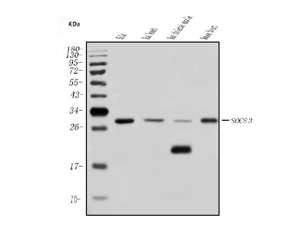
- Main image

- Experimental details
- Western blot analysis of SOCS3 using anti-SOCS3 antibody (A00274-2). Electrophoresis was performed on a 5-20% SDS-PAGE gel at 70V (Stacking gel) / 90V (Resolving gel) for 2-3 hours. The sample well of each lane was loaded with 50ug of sample under reducing conditions. Lane 1: human HELA whole cell lysates, Lane 2: rat heart tissue lysates, Lane 3: rat skeletal muscle tissue lysates, Lane 4: mouse heart tissue lysates. After Electrophoresis, proteins were transferred to a Nitrocellulose membrane at 150mA for 50-90 minutes. Blocked the membrane with 5% Non-fat Milk/ TBS for 1.5 hour at RT. The membrane was incubated with rabbit anti-SOCS3 antigen affinity purified polyclonal antibody (Catalog # A00274-2) at 0.5 μg/mL overnight at 4°C, then washed with TBS-0.1%Tween 3 times with 5 minutes each and probed with a goat anti-rabbit IgG-HRP secondary antibody at a dilution of 1:5000 for 1.5 hour at RT. The signal is developed using an Enhanced Chemiluminescent detection (ECL) kit (Catalog # EK1002) with Tanon 5200 system. A specific band was detected for SOCS3 at approximately 28-30KD. The expected band size for SOCS3 is at 28-30KD.
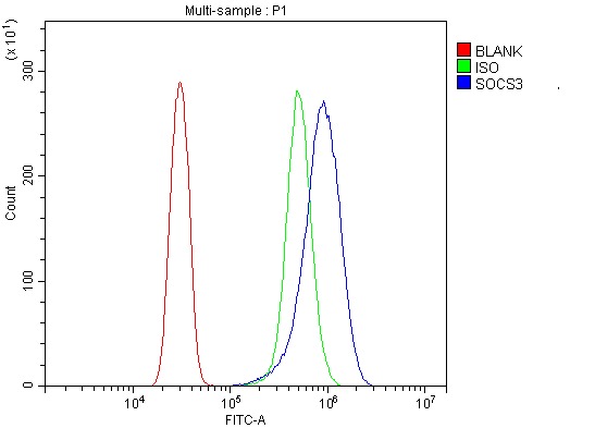
- Additional image