Explore
 Validate
 Learn
Learn Western blot
 Immunoprecipitation
Antibody data
- Antibody Data
- Antigen structure
- References [4]
- Comments [0]
- Validations
- Western blot [1]
- Immunocytochemistry [1]
Submit
Validation data
Reference
Comment
Report error
- Product number
- MA1-26249 - Provider product page

- Provider
- Invitrogen Antibodies
- Product name
- POLR2A Monoclonal Antibody (8WG16)
- Antibody type
- Monoclonal
- Antigen
- Purifed from natural sources
- Description
- MA1-26249 is expected to cross react with a wide range of other species due to sequence homology.
- Reactivity
- Human, Mouse, Bovine, Xenopus
- Host
- Mouse
- Isotype
- IgG
- Antibody clone number
- 8WG16
- Vial size
- 100 µg
- Concentration
- 1.1 mg/mL
- Storage
- Store at 4°C short term. For long term storage, store at -20°C, avoiding freeze/thaw cycles.
Submitted references RNA polymerase II clusters form in line with surface condensation on regulatory chromatin.
Enhancer Histone Acetylation Modulates Transcriptional Bursting Dynamics of Neuronal Activity-Inducible Genes.
Combining a Simple Method for DNA/RNA/Protein Co-Purification and Arabidopsis Protoplast Assay to Facilitate Viroid Research.
Transient RNA-DNA Hybrids Are Required for Efficient Double-Strand Break Repair.
Pancholi A, Klingberg T, Zhang W, Prizak R, Mamontova I, Noa A, Sobucki M, Kobitski AY, Nienhaus GU, Zaburdaev V, Hilbert L
Molecular systems biology 2021 Sep;17(9):e10272
Molecular systems biology 2021 Sep;17(9):e10272
Enhancer Histone Acetylation Modulates Transcriptional Bursting Dynamics of Neuronal Activity-Inducible Genes.
Chen LF, Lin YT, Gallegos DA, Hazlett MF, Gómez-Schiavon M, Yang MG, Kalmeta B, Zhou AS, Holtzman L, Gersbach CA, Grandl J, Buchler NE, West AE
Cell reports 2019 Jan 29;26(5):1174-1188.e5
Cell reports 2019 Jan 29;26(5):1174-1188.e5
Combining a Simple Method for DNA/RNA/Protein Co-Purification and Arabidopsis Protoplast Assay to Facilitate Viroid Research.
Jiang J, Ma J, Liu B, Wang Y
Viruses 2019 Apr 3;11(4)
Viruses 2019 Apr 3;11(4)
Transient RNA-DNA Hybrids Are Required for Efficient Double-Strand Break Repair.
Ohle C, Tesorero R, Schermann G, Dobrev N, Sinning I, Fischer T
Cell 2016 Nov 3;167(4):1001-1013.e7
Cell 2016 Nov 3;167(4):1001-1013.e7
No comments: Submit comment
Supportive validation
- Submitted by
- Invitrogen Antibodies (provider)
- Main image
- Experimental details
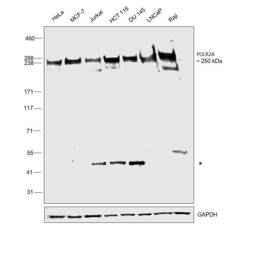
- Western blot was performed using Anti-POLR2A Monoclonal Antibody (8WG16) (Product # MA1-26249) and a 250 kDa band corresponding to POLR2A was observed along with uncharacterized bands (*) at 45 kDa across cell lines tested. Nuclear enriched extracts (40 µg lysate) of HeLa (Lane 1), MCF7 (Lane 2), Jurkat (Lane 3), HCT 116 (Lane 4), DU 145 (Lane 5), LNCaP (Lane 6) and Raji (Lane 7) were electrophoresed using NuPAGE™ 3-8% Tris-Acetate Protein Gel (Product # EA0378BOX). Resolved proteins were equilibrated with 20% ethanol and then transferred onto a nitrocellulose membrane (Product # IB23002) by iBlot® 2 Dry Blotting System (Product # IB21001). The blot was probed with the primary antibody (1:1000 dilution) and detected by chemiluminescence with Goat anti-Mouse IgG (H+L) Superclonal™ Recombinant Secondary Antibody, HRP (Product # A28177,1:20000 dilution using the iBright™ FL1500 Imaging System (Product # A44115). Chemiluminescent detection was performed using SuperSignal™ West Pico PLUS Chemiluminescent Substrate (Product # 34580).
Supportive validation
- Submitted by
- Invitrogen Antibodies (provider)
- Main image

- Experimental details
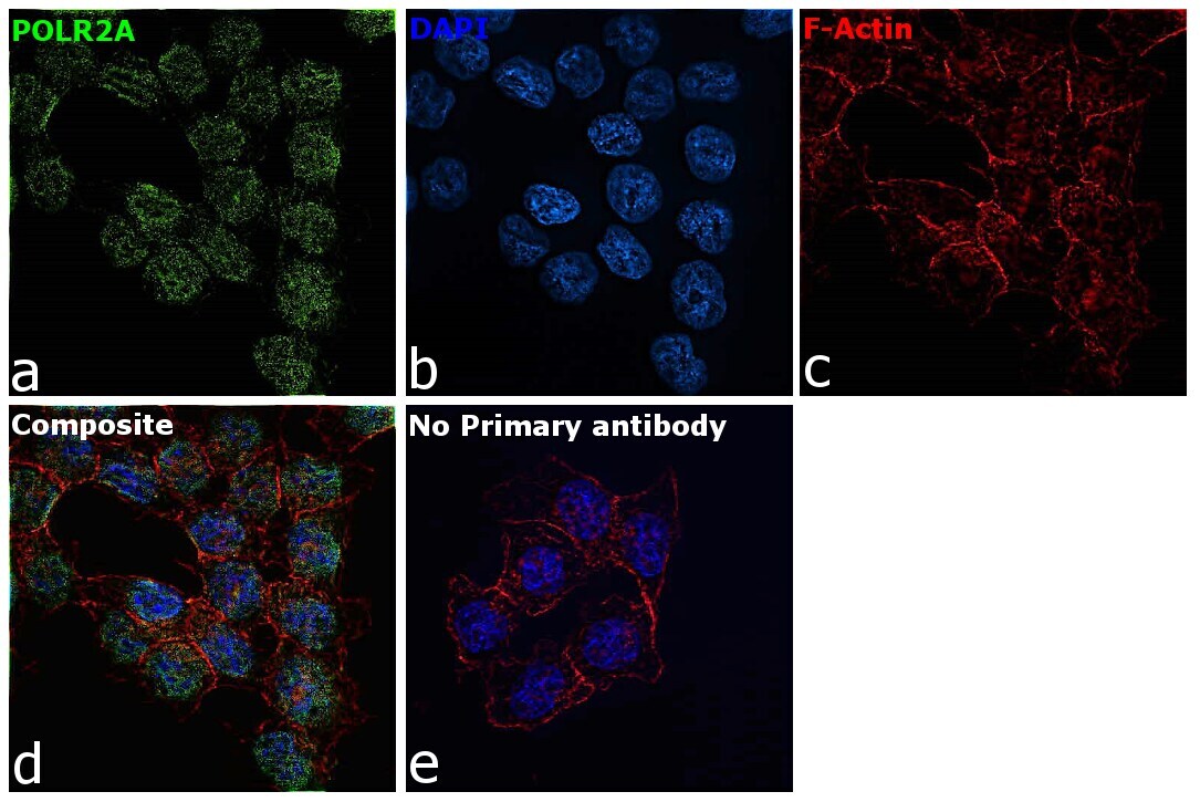
- Immunofluorescence analysis of POLR2A Monoclonal Antibody (8WG16) was performed using 70% confluent log phase HCT 116 cells. The cells were fixed with 4% paraformaldehyde for 10 minutes, permeabilized with 0.1% Triton™ X-100 for 15 minutes, and blocked with 2% BSA for 45 minutes at room temperature. The cells were labeled with POLR2A Monoclonal Antibody (8WG16) (Product # MA1-26249) at 1:100 dilution in 0.1% BSA, incubated at 4 degree celsius overnight and then labeled with Donkey anti-Mouse IgG (H+L) Highly Cross-Adsorbed Secondary Antibody, Alexa Fluor Plus 488 (Product # A32766), (1:2000 dilution), for 45 minutes at room temperature (Panel a: Green). Nuclei (Panel b:Blue) were stained with ProLong™ Diamond Antifade Mountant with DAPI (Product # P36962). F-actin (Panel c: Red) was stained with Rhodamine Phalloidin (Product # R415, 1:300 dilution). Panel d represents the merged image showing Nuclear localization. Panel e represents control cells with no primary antibody to assess background. The images were captured at 60X magnification.