 Explore
 Validate
 Learn
Learn Western blot
Antibody data
- Antibody Data
- Antigen structure
- References [1]
- Comments [0]
- Validations
- Western blot [1]
- Immunocytochemistry [1]
- Chromatin Immunoprecipitation [1]
Submit
Validation data
Reference
Comment
Report error
- Product number
- MA1-10882 - Provider product page

- Provider
- Invitrogen Antibodies
- Product name
- POLR2A Monoclonal Antibody (8WG16)
- Antibody type
- Monoclonal
- Antigen
- Purifed from natural sources
- Description
- MA1-10882 detects RNA Polymerase II from eukaryotic samples.
- Antibody clone number
- 8WG16
- Concentration
- 1.1 mg/mL
Submitted references S100A14: novel modulator of terminal differentiation in esophageal cancer.
Chen H, Ma J, Sunkel B, Luo A, Ding F, Li Y, He H, Zhang S, Xu C, Jin Q, Wang Q, Liu Z
Molecular cancer research : MCR 2013 Dec;11(12):1542-53
Molecular cancer research : MCR 2013 Dec;11(12):1542-53
No comments: Submit comment
Supportive validation
- Submitted by
- Invitrogen Antibodies (provider)
- Main image

- Experimental details
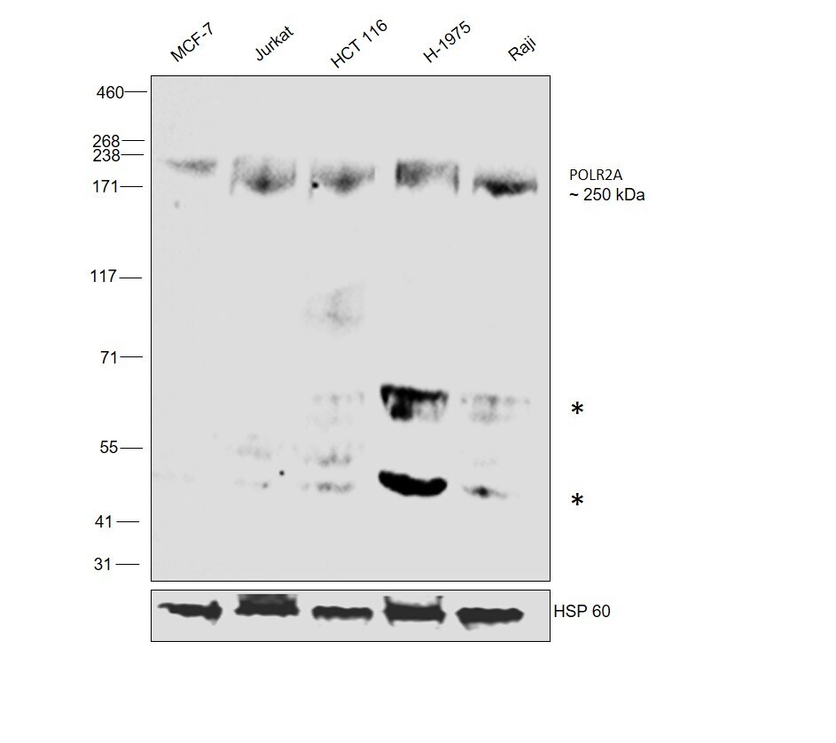
- Western blot was performed using Anti-POLR2A Monoclonal Antibody (8WG16) (Product # MA1-10882) and a 250 kDa bands along with uncharacterized bands (*) corresponding to POLR2A was observed across cell lines tested. Nuclear enriched extracts (40 µg lysate) of MCF7 (Lane 1), Jurkat (Lane 2), HCT 116 (Lane 3), H-1975 (Lane 4) and Raji (Lane 5) were electrophoresed using NuPAGE™ 3-8% Tris-Acetate Protein Gel (Product # EA0378BOX). Resolved proteins were equilibrated with 20% ethanol then transferred onto a nitrocellulose membrane (Product # IB23002) by iBlot® 2 Dry Blotting System (Product # IB21001). The blot was probed with the primary antibody (1 µg/mL) and detected by chemiluminescence with Goat anti-Mouse IgG (H+L) Superclonal™ Recombinant Secondary Antibody, HRP (Product # A28177,1:20000 dilution using the iBright™ FL1500 Imaging System (Product # A44115). Chemiluminescent detection was performed using SuperSignal™ West Pico PLUS Chemiluminescent Substrate (Product # 34580).
Supportive validation
- Submitted by
- Invitrogen Antibodies (provider)
- Main image

- Experimental details
- Immunofluorescence analysis of POLR2A Monoclonal Antibody (8WG16) was performed using 70% confluent log phase HCT 116 cells. The cells were fixed with 4% paraformaldehyde for 10 minutes, permeabilized with 0.1% Triton™ X-100 for 15 minutes, and blocked with 2% BSA for 45 minutes at room temperature. The cells were labeled with POLR2A Monoclonal Antibody (8WG16) (Product # MA1-10882) at 5 µg/mL in 0.1% BSA, incubated at 4 degree celsius overnight and then labeled with Donkey anti-Mouse IgG (H+L) Highly Cross-Adsorbed Secondary Antibody, Alexa Fluor Plus 488 (Product # A32766), (1:2000 dilution), for 45 minutes at room temperature (Panel a: Green). Nuclei (Panel b:Blue) were stained with ProLong™ Diamond Antifade Mountant with DAPI (Product # P36962). F-actin (Panel c: Red) was stained with Rhodamine Phalloidin (Product # R415, 1:300 dilution). Panel d represents the merged image showing Nuclear localization. Panel e represents control cells with no primary antibody to assess background. The images were captured at 60X magnification.

Supportive validation
- Submitted by
- Invitrogen Antibodies (provider)
- Main image
- Experimental details
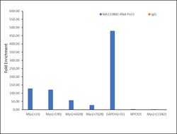
- Chromatin Immunoprecipitation (ChIP) was performed using POLR2A Monoclonal Antibody (8WG16) (Product # MA1-10882, 5 µg) on sheared chromatin from a million HCT 116 cells using the MAGnify ChIP System (Product # 49-2024). Normal Mouse IgG was used as a negative IP control. The purified DNA was analyzed by qPCR with PCR primer pairs over Myc and GAPDH genes (active), and MYOD1 and Myc (+11382) (inactive). Antibody specificity was demonstrated by detection of enrichment of the target protein at specific gene loci. Data is presented as fold enrichment of the antibody signal versus the Mouse Isotype using the comparative CT method.
