 Explore
 Validate
 Learn
Learn Western blot
 Immunocytochemistry
 Flow cytometry
Antibody data
- Antibody Data
- Antigen structure
- References [5]
- Comments [0]
- Validations
- Immunocytochemistry [2]
- Other assay [3]
Submit
Validation data
Reference
Comment
Report error
- Product number
- 459000 - Provider product page

- Provider
- Invitrogen Antibodies
- Product name
- ATP5H Monoclonal Antibody (7F9BG1)
- Antibody type
- Monoclonal
- Antigen
- Other
- Description
- For mouse and rat samples, this antibody will only recognize ATP5H in purified mitochondrial samples. Mouse and rat cell lysates and tissue homogenates are not recommended with this antibody. For positive control, use Isolated mitochondria from Human heart, Bovine heart, Rat heart, Mouse heart, and HepG2, Cultured Human embryonic lung-derived fibroblasts (strain MRC5), HeLa cells.
- Reactivity
- Human, Mouse, Rat, Bovine
- Host
- Mouse
- Isotype
- IgG
- Antibody clone number
- 7F9BG1
- Vial size
- 100 μg
- Concentration
- 1 mg/mL
- Storage
- 4°C, do not freeze
Submitted references ATM-mediated mitochondrial damage response triggered by nuclear DNA damage in normal human lung fibroblasts.
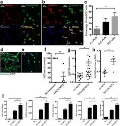
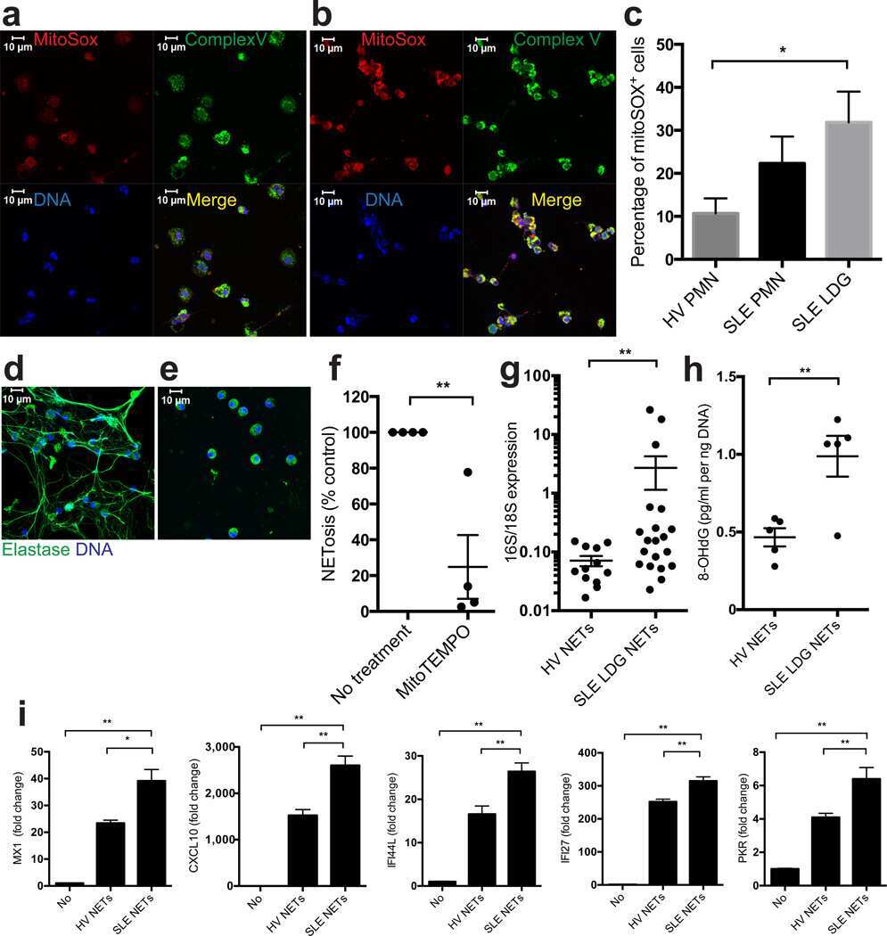
Neutrophil extracellular traps enriched in oxidized mitochondrial DNA are interferogenic and contribute to lupus-like disease.
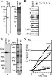
Assembly of human mitochondrial ATP synthase through two separate intermediates, F1-c-ring and b-e-g complex.
Population of ATP synthase molecules in mitochondria is limited by available 6.8-kDa proteolipid protein (MLQ).
Tau oligomers impair memory and induce synaptic and mitochondrial dysfunction in wild-type mice.
Shimura T, Sasatani M, Kawai H, Kamiya K, Kobayashi J, Komatsu K, Kunugita N
Cell cycle (Georgetown, Tex.) 2017;16(24):2345-2354
Cell cycle (Georgetown, Tex.) 2017;16(24):2345-2354
Neutrophil extracellular traps enriched in oxidized mitochondrial DNA are interferogenic and contribute to lupus-like disease.
Lood C, Blanco LP, Purmalek MM, Carmona-Rivera C, De Ravin SS, Smith CK, Malech HL, Ledbetter JA, Elkon KB, Kaplan MJ
Nature medicine 2016 Feb;22(2):146-53
Nature medicine 2016 Feb;22(2):146-53
Assembly of human mitochondrial ATP synthase through two separate intermediates, F1-c-ring and b-e-g complex.
Fujikawa M, Sugawara K, Tanabe T, Yoshida M
FEBS letters 2015 Sep 14;589(19 Pt B):2707-12
FEBS letters 2015 Sep 14;589(19 Pt B):2707-12
Population of ATP synthase molecules in mitochondria is limited by available 6.8-kDa proteolipid protein (MLQ).
Fujikawa M, Ohsakaya S, Sugawara K, Yoshida M
Genes to cells : devoted to molecular & cellular mechanisms 2014 Feb;19(2):153-60
Genes to cells : devoted to molecular & cellular mechanisms 2014 Feb;19(2):153-60
Tau oligomers impair memory and induce synaptic and mitochondrial dysfunction in wild-type mice.
Lasagna-Reeves CA, Castillo-Carranza DL, Sengupta U, Clos AL, Jackson GR, Kayed R
Molecular neurodegeneration 2011 Jun 6;6:39
Molecular neurodegeneration 2011 Jun 6;6:39
No comments: Submit comment
Supportive validation
- Submitted by
- Invitrogen Antibodies (provider)
- Main image

- Experimental details
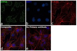
- Immunofluorescence analysis of ATP5H was performed using Hep G2 cells. The cells were fixed with 4% paraformaldehyde for 10 minutes, permeabilized with 0.1% Triton™ X-100 for 15 minutes, and blocked with 2% BSA for 1 hour at room temperature. The cells were labeled with ATP5H Mouse Monoclonal Antibody (Product # 459000) at 5 µg/mL in 0.1% BSA and incubated overnight at 4 degree and then labeled with Goat anti-Mouse IgG (H+L) Highly Cross-Adsorbed Secondary Antibody, Alexa Fluor Plus 488 (Product # A32723) at a dilution of 1:2000 for 45 minutes at room temperature (Panel a: green). Nuclei (Panel b: blue) were stained with ProLong™ Diamond Antifade Mountant with DAPI (Product # P36962). F-actin (Panel c: red) was stained with Rhodamine Phalloidin (Product # R415, 1:300). Panel d represents the composite image showing mitochondrial pattern of ATP5H. Panel e represents control cells with no primary antibody to assess background. The images were captured at 60X magnification.

- Submitted by
- Invitrogen Antibodies (provider)
- Main image
- Experimental details
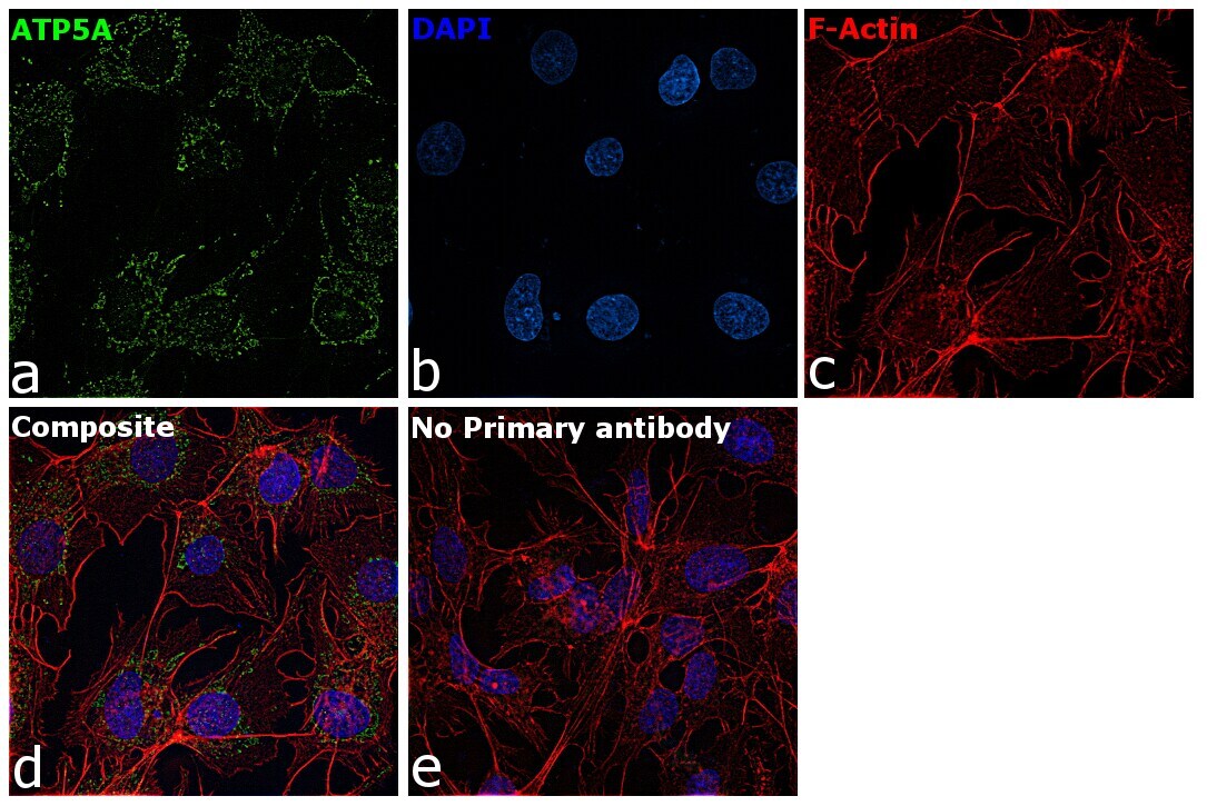
- Immunofluorescence analysis of ATP5H was performed using Hep G2 cells. The cells were fixed with 4% paraformaldehyde for 10 minutes, permeabilized with 0.1% Triton™ X-100 for 15 minutes, and blocked with 2% BSA for 1 hour at room temperature. The cells were labeled with ATP5H Mouse Monoclonal Antibody (Product # 459000) at 5 µg/mL in 0.1% BSA and incubated overnight at 4 degree and then labeled with Goat anti-Mouse IgG (H+L) Highly Cross-Adsorbed Secondary Antibody, Alexa Fluor Plus 488 (Product # A32723) at a dilution of 1:2000 for 45 minutes at room temperature (Panel a: green). Nuclei (Panel b: blue) were stained with ProLong™ Diamond Antifade Mountant with DAPI (Product # P36962). F-actin (Panel c: red) was stained with Rhodamine Phalloidin (Product # R415, 1:300). Panel d represents the composite image showing mitochondrial pattern of ATP5H. Panel e represents control cells with no primary antibody to assess background. The images were captured at 60X magnification.
Supportive validation
- Submitted by
- Invitrogen Antibodies (provider)
- Main image
- Experimental details
- NULL
- Submitted by
- Invitrogen Antibodies (provider)
- Main image
- Experimental details
- NULL

- Submitted by
- Invitrogen Antibodies (provider)
- Main image
- Experimental details
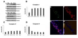
- Figure 6 Tau oligomers induce mitochondrial alterations . (A) Representative western blot of mouse brain homogenate. The levels of complex I, complex V, and caspase-9 were measured by band quantification and normalized with the levels of tubulin. Abbreviations are as in Fig. 4. (B) Complex I levels were significantly lower in the hemisphere injected with tau oligomers in comparison with the ones injected with fibrils or monomers. (C) No differences in the levels of complex V were observed in any of the groups. (D) Caspase-9 activation was significantly higher in tau oligomer groups over both tau fibril- and monomer-injected groups. (E-G) Double staining between human tau-specific antibody HT7 (green fluorescence) and mitochondrial porin antibody (red fluorescence) demonstrates the internalization of tau oligomers in CA1 cells and their interaction with the mitochondria. (H) The co-localization of tau oligomers with mitochondria was confirmed by ICA of the signal. Data are represented as mean +- SE. *p < 0.01, n = 6.
