Explore
 Validate
 Learn
Learn Western blot
 ELISA
ELISAAntibody data
- Antibody Data
- Antigen structure
- References [1]
- Comments [0]
- Validations
- Western blot [2]
- Immunohistochemistry [1]
Submit
Validation data
Reference
Comment
Report error
- Product number
- NBP1-51678 - Provider product page

- Provider
- Novus Biologicals
- Proper citation
- Novus Cat#NBP1-51678, RRID:AB_11019427
- Product name
- Mouse Monoclonal Lysine (K)-specific Demethylase 3A/KDM3A/JMJD1A Antibody
- Antibody type
- Monoclonal
- Description
- Ammonium sulfate precipitation.
- Reactivity
- Human
- Host
- Mouse
- Isotype
- IgG
- Vial size
- 0.1 ml
- Concentration
- 1.0 mg/ml
- Storage
- Store at 4C short term. Aliquot and store at -20C long term. Avoid freeze-thaw cycles.
Submitted references Epigenetic regulation of mouse sex determination by the histone demethylase Jmjd1a.
Kuroki S, Matoba S, Akiyoshi M, Matsumura Y, Miyachi H, Mise N, Abe K, Ogura A, Wilhelm D, Koopman P, Nozaki M, Kanai Y, Shinkai Y, Tachibana M
Science (New York, N.Y.) 2013 Sep 6;341(6150):1106-9
Science (New York, N.Y.) 2013 Sep 6;341(6150):1106-9
No comments: Submit comment
Supportive validation
- Submitted by
- Novus Biologicals (provider)
- Main image

- Experimental details
- Simple Western: Lysine (K)-specific Demethylase 3A/KDM3A/JMJD1A Antibody (1E12) [NBP1-51678] - Simple Western lane view shows a specific band for JMJD1A in 0.5 mg/ml of HepG2 lysate. This experiment was performed under reducing conditions using the 12-230 kDa separation system.

- Submitted by
- Novus Biologicals (provider)
- Main image

- Experimental details
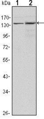
- Western Blot: Lysine (K)-specific Demethylase 3A/KDM3A/JMJD1A Antibody (1E12) [NBP1-51678] - Western blot analysis using JMJD1A mouse mAb against Hela (1) and HepG2 (2) cell lysates.
Supportive validation
- Submitted by
- Novus Biologicals (provider)
- Main image

- Experimental details
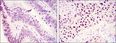
- Immunohistochemistry-Paraffin: Lysine (K)-specific Demethylase 3A/KDM3A/JMJD1A Antibody (1E12) [NBP1-51678] - Immunohistochemical analysis of paraffin-embedded colonic cancer tissues (left) and lung cancer tissues (right) using JMJD1A mouse mAb with DAB staining.