Explore
 Validate
 Learn
Learn Western blot
Antibody data
- Antibody Data
- Antigen structure
- References [1]
- Comments [0]
- Validations
- Western blot [7]
- ELISA [2]
Submit
Validation data
Reference
Comment
Report error
- Product number
- GTX106585 - Provider product page

- Provider
- GeneTex
- Proper citation
- GeneTex Cat#GTX106585, RRID:AB_1950503
- Product name
- HN1 antibody
- Antibody type
- Polyclonal
- Reactivity
- Human
- Host
- Rabbit
Submitted references MiR-132 prohibits proliferation, invasion, migration, and metastasis in breast cancer by targeting HN1
Zhang Z, Chen W, Wu Y, Liang H, Zhang B
Biochemical and Biophysical Research Communications 2014 November;454(1):109-114
Biochemical and Biophysical Research Communications 2014 November;454(1):109-114
No comments: Submit comment
Supportive validation
- Submitted by
- GeneTex (provider)
- Main image
- Experimental details
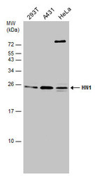
- Sample(30 ug whole cell lysate)A:A431(GTX27909)B:H1299C:HeLa S3(GTX14654)D:Hep G2 (GTX27900)12% SDS PAGEGTX106585 diluted at 1:1000
- Validation comment
- WB
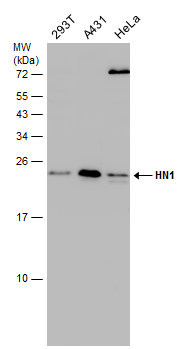
- Submitted by
- GeneTex (provider)
- Main image
- Experimental details
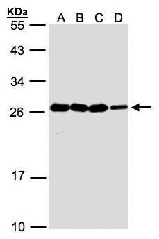
- Various whole cell extracts (30 ?g) were separated by 15% SDS-PAGE, and the membrane was blotted with HN1 antibody (GTX106585) diluted at 1:500. The HRP-conjugated anti-rabbit IgG antibody (GTX213110-01) was used to detect the primary antibody.
- Submitted by
- GeneTex (provider)
- Main image
- Experimental details
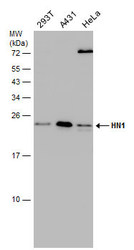
- Various whole cell extracts (30 ?g) were separated by 15% SDS-PAGE, and the membrane was blotted with HN1 antibody (GTX106585) diluted at 1:500. The HRP-conjugated anti-rabbit IgG antibody (GTX213110-01) was used to detect the primary antibody.
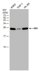
- Submitted by
- GeneTex (provider)
- Main image
- Experimental details
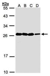
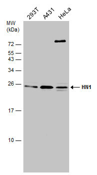
- Various whole cell extracts (30 ?g) were separated by 15% SDS-PAGE, and the membrane was blotted with HN1 antibody (GTX106585) diluted at 1:3000.
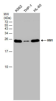
- Submitted by
- GeneTex (provider)
- Main image
- Experimental details
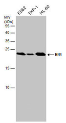
- Various whole cell extracts (30 ?g) were separated by 15% SDS-PAGE, and the membrane was blotted with HN1 antibody (GTX106585) diluted at 1:3000.
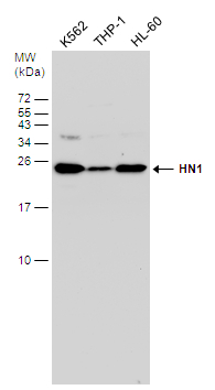
- Submitted by
- GeneTex (provider)
- Main image

- Experimental details
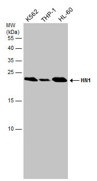
- Various whole cell extracts (30 ?g) were separated by 15% SDS-PAGE, and the membrane was blotted with HN1 antibody (GTX106585) diluted at 1:3000.
- Submitted by
- GeneTex (provider)
- Main image
- Experimental details
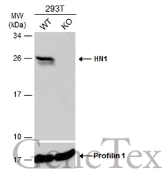
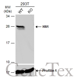
- Wild-type (WT) and HN1 knockout (KO) 293T cell extracts (30 ?g) were separated by 12% SDS-PAGE, and the membrane was blotted with HN1 antibody (GTX106585) diluted at 1:1000. The HRP-conjugated anti-rabbit IgG antibody (GTX213110-01) was used to detect the primary antibody.
Supportive validation
- Submitted by
- GeneTex (provider)
- Main image

- Experimental details
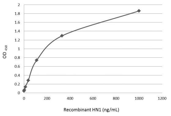
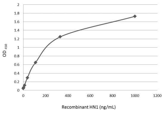
- An ELISA plate is coated with 50 ?L of HN1 recombinant protein at concentration ranged from 0.004 ?g/mL to 1 ?g/mL. The coated protein is detected with HN1 antibody (GTX106585) at 0.25 ?g/mL.
- Submitted by
- GeneTex (provider)
- Main image
- Experimental details
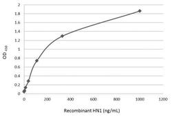
- An ELISA plate is coated with 50 ?L of HN1 recombinant protein at concentration ranged from 0.004 ?g/mL to 1 ?g/mL. The coated protein is detected with HN1 antibody (GTX106585) at 0.25 ?g/mL.