Explore
 Validate
 Learn
Learn Western blot
 Immunocytochemistry
Antibody data
- Antibody Data
- Antigen structure
- References [4]
- Comments [0]
- Validations
- Immunocytochemistry [2]
- Immunohistochemistry [1]
- Other assay [1]
Submit
Validation data
Reference
Comment
Report error
- Product number
- PA5-35201 - Provider product page

- Provider
- Invitrogen Antibodies
- Product name
- ATG5 Polyclonal Antibody
- Antibody type
- Polyclonal
- Antigen
- Synthetic peptide
- Description
- This antibody is predicted to react with bovine, porcine and rat based on sequence homology.
- Reactivity
- Human, Mouse
- Host
- Rabbit
- Isotype
- IgG
- Vial size
- 400 μL
- Concentration
- 0.5 mg/mL
- Storage
- Store at 4°C short term. For long term storage, store at -20°C, avoiding freeze/thaw cycles.
Submitted references Knockdown of VDAC1 alleviates the cognitive dysfunction secondary to sepsis-associated encephalopathy.
Autophagy core protein ATG5 is required for elongating spermatid development, sperm individualization and normal fertility in male mice.
Mechanistic studies of cytotoxic activity of the mesoionic compound MIH 2.4Bl in MCF-7 breast cancer cells.
Aggresome-Like Formation Promotes Resistance to Proteotoxicity in Cells from Long-Lived Species.
Cai M, Du B, Si Y, Miao J, Ge J, Zhang J, Song J, Bao H
American journal of translational research 2021;13(7):7538-7555
American journal of translational research 2021;13(7):7538-7555
Autophagy core protein ATG5 is required for elongating spermatid development, sperm individualization and normal fertility in male mice.
Huang Q, Liu Y, Zhang S, Yap YT, Li W, Zhang D, Gardner A, Zhang L, Song S, Hess RA, Zhang Z
Autophagy 2021 Jul;17(7):1753-1767
Autophagy 2021 Jul;17(7):1753-1767
Mechanistic studies of cytotoxic activity of the mesoionic compound MIH 2.4Bl in MCF-7 breast cancer cells.
de Mascena Costa LA, Debnath D, Harmon AC, de Sousa Araújo S, da Silva Souza HD, de Athayde Filho PF, Wischral A, Adrião Gomes Filho M, Mathis JM
Oncology letters 2020 Sep;20(3):2291-2301
Oncology letters 2020 Sep;20(3):2291-2301
Aggresome-Like Formation Promotes Resistance to Proteotoxicity in Cells from Long-Lived Species.
Sunchu B, Riordan RT, Yu Z, Almog I, Dimas-Munoz J, Drake AC, Perez VI
The journals of gerontology. Series A, Biological sciences and medical sciences 2020 Jul 13;75(8):1439-1447
The journals of gerontology. Series A, Biological sciences and medical sciences 2020 Jul 13;75(8):1439-1447
No comments: Submit comment
Supportive validation
- Submitted by
- Invitrogen Antibodies (provider)
- Main image

- Experimental details
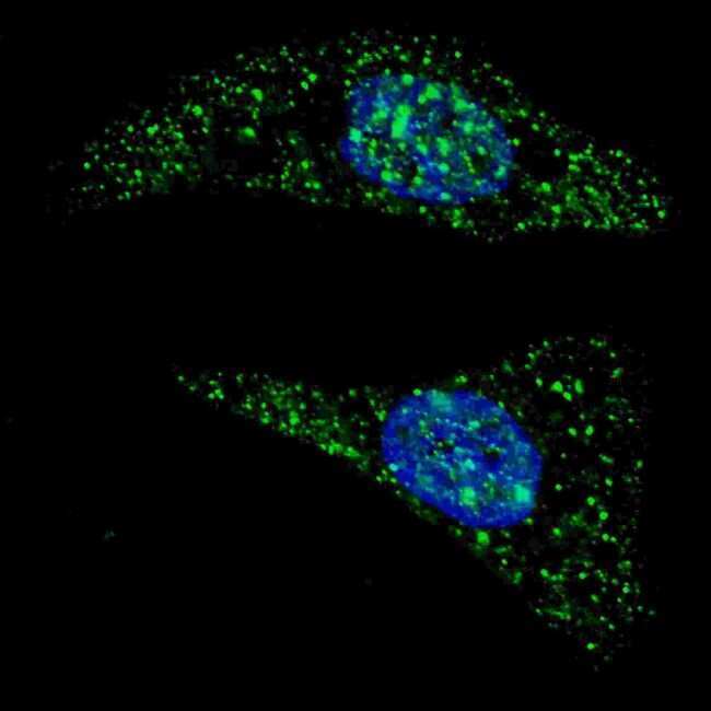
- Immunofluorescent analysis of ATG5 showing staining in the cytoplasm of U-251 cells. U251 cells were treated with Chloroquine (50uM, 16h), fixed with 4% PFA (20 min) and permeabilized with Triton X-100 (0.2%, 30 min). Cells were probed with an ATG5 polyclonal antibody (Product # PA5-35201) (1:100, 2h at room temperature) followed by detection using a fluorescent conjugated secondary antibody (green) (1:1000, 1h). Nuclei were stained with Hoechst 33342 (blue) (10 µg/mL, 5 min).
- Submitted by
- Invitrogen Antibodies (provider)
- Main image

- Experimental details
- Immunocytochemistry analysis of ATG5 in U251 cells. Samples were incubated with ATG5 polyclonal antibody (Product # PA5-35201) using a dilution of 1:200 for 2 hr at room temperature followed by Alexa Fluor® 488 conjugated donkey anti-rabbit antibody (green) at a dilution of 1:1,000 for 1hr. Cells were treated with Chloroquine (50 μM, 16hr), then fixed with 4% PFA (20 min), and permeabilized with Triton X-100 (0.2%, 30 min). Nuclei were counterstained with Hoechst 33342 (blue) (10 µg/mL, 5 min). ATG5 immunoreactivity is localized to autophagic vacuoles in the cytoplasm of U251 cells.

Supportive validation
- Submitted by
- Invitrogen Antibodies (provider)
- Main image
- Experimental details
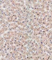
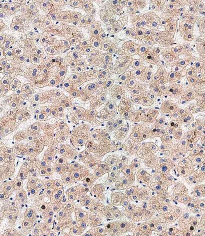
- Immunohistochemistry analysis of ATG5 in paraffin-embedded human liver tissue. Samples were incubated with ATG5 polyclonal antibody (Product # PA5-35201) using a dilution of 1:500 for 1 hour at room temperature followed by an undiluted biotinylated CRF Anti-Polyvalent HRP Polymer antibody. Tissue was fixed with formaldehyde at room temperature, antigen retrieval was by heat mediation with a EDTA buffer (pH 9.0).
Supportive validation
- Submitted by
- Invitrogen Antibodies (provider)
- Main image

- Experimental details
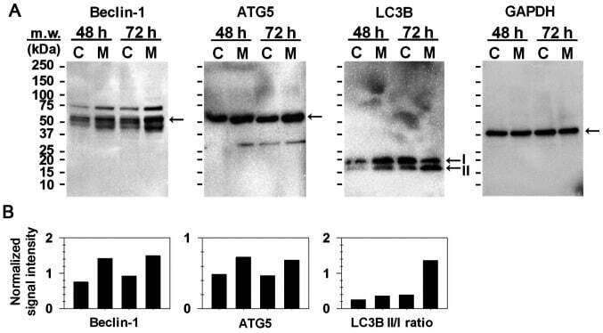
- Figure 2. Effect of MIH 2.4Bl on the induction of autophagy in MCF-7 cells. (A) Expression of Beclin-1, ATG5, LC3B II/I and GAPDH were determined by western blot analysis in control or MIH 2.4Bl treated cells after 48 or 72 h of culture. (B) Densitometry analysis of Beclin-1, ATG5, LC3B II and LC3B I expression was performed and normalized to GAPDH. Data are presented as histograms of normalized Beclin-1 and ATG5 intensity values, and the intensity ratios of LC3B-II to LC3B-I. LC3B, microtubule-associated protein 1A/1B light chain 3B. C, control cells; M, MIH 2.4Bl treated cells.