Explore
 Validate
 Learn
LearnGTX48583
antibody from GeneTex
Targeting: ATG5
APG5, APG5L, ASP, hAPG5
 Western blot Immunocytochemistry
 Immunoprecipitation Immunohistochemistry Flow cytometry Proximity ligation assay
Antibody data
- Antibody Data
- Antigen structure
- References [1]
- Comments [0]
- Validations
- Western blot [1]
- Immunocytochemistry [1]
- Immunohistochemistry [2]
Submit
Validation data
Reference
Comment
Report error
- Product number
- GTX48583 - Provider product page

- Provider
- GeneTex
- Proper citation
- GeneTex Cat#GTX48583, RRID:AB_10620228
- Product name
- ATG5 antibody
- Antibody type
- Polyclonal
- Reactivity
- Human, Mouse, Rat, Bovine, Porcine, Simian, Xenopus, Zebrafish
- Host
- Rabbit
Submitted references Autophagy-mediated degradation of nuclear envelope proteins during oncogene-induced senescence.
Lenain C, Gusyatiner O, Douma S, van den Broek B, Peeper DS
Carcinogenesis 2015 Nov;36(11):1263-74
Carcinogenesis 2015 Nov;36(11):1263-74
No comments: Submit comment
Supportive validation
- Submitted by
- GeneTex (provider)
- Main image
- Experimental details
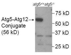
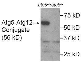
- Western Blot: Detection of ATG5 in mouse wildtype ES cell lysate (Lane 1) using GTX48583. Lane 2 is a mouse ATG5 KO ES cell lysate (negative control). Atg5-/- ES cells from Dr. Noboru Mizushima [Mizushima, N. et al. J. Cell Biol. 152 (2001)] Photo courtesy of Dr. Beth Levine, UT Southwestern Medical Center"
Supportive validation
- Submitted by
- GeneTex (provider)
- Main image
- Experimental details
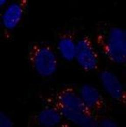
- Immunocytochemistry/Immunofluorescence: Immunofluorescent staining of SY5Y cells using GTX48583 at 1:250. Incubated overnight at 4 degrees. Photo courtesy of an anonymous collaborator. Immunofluorescent staining of SY5Y cells using GTX48583 at 1:250. Incubated overnight at 4 degrees. Photo courtesy of an anonymous collaborator.

Supportive validation
- Submitted by
- GeneTex (provider)
- Main image

- Experimental details
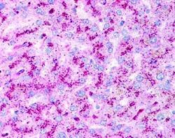
- Immunohistochemistry: Staining of hepatocytes using GTX48583 at 2.5μg/ml. Human liver hepatocytes 40X magnification.
- Submitted by
- GeneTex (provider)
- Main image
- Experimental details
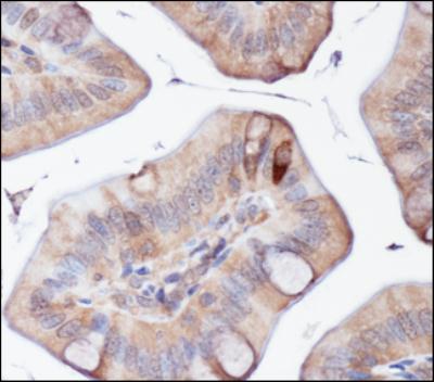
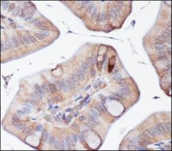
- Immunohistochemistry: ATG5 Antibody [GTX48583] - Analysis of ATG5 in mouse intestine using DAB with hematoxylin counterstain.