Explore
 Validate
 Learn
Learn Western blot
 ELISA
ELISA Immunocytochemistry
Antibody data
- Antibody Data
- Antigen structure
- References [1]
- Comments [0]
- Validations
- Immunocytochemistry [2]
- Flow cytometry [2]
- Other assay [1]
Submit
Validation data
Reference
Comment
Report error
- Product number
- MA5-38452 - Provider product page

- Provider
- Invitrogen Antibodies
- Product name
- ATG5 Monoclonal Antibody (8E8G6)
- Antibody type
- Monoclonal
- Antigen
- Synthetic peptide
- Description
- This antibody has been tested in indirect ELISA.
- Reactivity
- Human
- Host
- Mouse
- Isotype
- IgG
- Antibody clone number
- 8E8G6
- Vial size
- 100 µg
- Concentration
- 1 mg/mL
- Storage
- Store at 4°C short term. For long term storage, store at -20°C, avoiding freeze/thaw cycles.
Submitted references AMPK modulation ameliorates dominant disease phenotypes of CTRP5 variant in retinal degeneration.
Miyagishima KJ, Sharma R, Nimmagadda M, Clore-Gronenborn K, Qureshy Z, Ortolan D, Bose D, Farnoodian M, Zhang C, Fausey A, Sergeev YV, Abu-Asab M, Jun B, Do KV, Kautzman Guerin MA, Calandria J, George A, Guan B, Wan Q, Sharp RC, Cukras C, Sieving PA, Hufnagel RB, Bazan NG, Boesze-Battaglia K, Miller S, Bharti K
Communications biology 2021 Dec 9;4(1):1360
Communications biology 2021 Dec 9;4(1):1360
No comments: Submit comment
Supportive validation
- Submitted by
- Invitrogen Antibodies (provider)
- Main image

- Experimental details
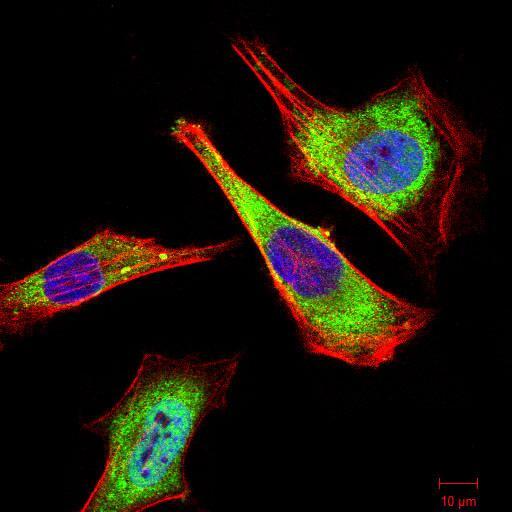
- Immunocytochemistry/Immunofluorescence analysis of ATG5 in HeLa cells using ATG5 Monoclonal Antibody (Product # MA5-38452) (green). Blue: DRAQ5 fluorescent DNA dye. Red: Actin filaments have been labeled with Alexa Fluor- 555 phalloidin. Secondary antibody from Fisher (Product # 35503)

- Submitted by
- Invitrogen Antibodies (provider)
- Main image

- Experimental details
- Immunocytochemistry/Immunofluorescence analysis of ATG5 in HeLa cells using ATG5 Monoclonal Antibody (Product # MA5-38452) (green). Blue: DRAQ5 fluorescent DNA dye. Red: Actin filaments have been labeled with Alexa Fluor- 555 phalloidin. Secondary antibody from Fisher (Product # 35503)
Supportive validation
- Submitted by
- Invitrogen Antibodies (provider)
- Main image

- Experimental details
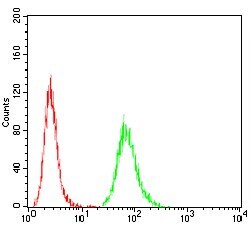
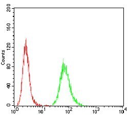
- Flow Cytometry analysis of ATG5 in HeLa cells using ATG5 Monoclonal Antibody (Product # MA5-38452) (green) and negative control (red).
- Submitted by
- Invitrogen Antibodies (provider)
- Main image
- Experimental details
- Flow Cytometry analysis of ATG5 in HeLa cells using ATG5 Monoclonal Antibody (Product # MA5-38452) (green) and negative control (red).

Supportive validation
- Submitted by
- Invitrogen Antibodies (provider)
- Main image
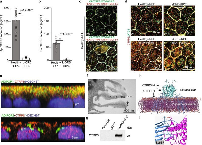
- Experimental details
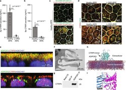
- Fig. 2 Expression and localization of CTRP5 in L-ORD-iRPE. a , b Apical and basal CTRP5 secretion measured by ELISA in the culture medium are both significantly decreased ( n = 14). c Coexpression of V5-tagged WT CTRP5 (green) and FLAG-tagged S163R CTRP5 (red) in healthy-iRPE. V5-tagged WT CTRP5 expressing lentivirus construct was transduced at MOI 0.5 for both top and bottom panels. MOI of Flag-tagged S163R CTRP5 expressing lentivirus construct was 0.5 for cells in the top panel and 3.0 for cells in the bottom panel. Scale bar: 10 um d Confocal microscopy images of untreated and bafilomycin (BafA1) treated (3 h) healthy-iRPE and L-ORD-iRPE co-stained with CTRP5 (red) and ATG5 (green). ( n = 3 images per condition). Scale bar: 10 um. e Representative confocal microscopy images showing colocalization of CTRP5 (red) with membrane receptors ADIPOR1 (green, upper panel) and no colocalization with ADIPOR2 (green, lower panel). Nuclear stain (blue). Scale bar: 5 um. f TEM image of immunogold labeled ADIPOR1 (6 nm gold particle) and CTRP5 (12 nm gold particle) demonstrate the co-binding of two proteins (arrow). Scale bar: 500 nm. g Western blot detects CTRP5 in the membrane fraction of iRPE cells immunoprecipitated using anti-ADIPOR1 antibodies and not in lanes immunoprecipitated with the IgG antibody or with only beads. h Probabilistic model of the interaction of integral membrane protein ADIPOR1 (blue) and CTRP5 (teal) determined using published crystallographic structures and ref