 Explore
 Validate
 Learn
Learn25625-1-AP
antibody from Proteintech Group
Targeting: CHCHD3
FLJ20420, Mic19, MICOS19, MINOS3, PPP1R22
 Western blot
 ELISA
ELISAAntibody data
- Antibody Data
- Antigen structure
- References [42]
- Comments [0]
- Validations
- Western blot [3]
Submit
Validation data
Reference
Comment
Report error
- Product number
- 25625-1-AP - Provider product page

- Provider
- Proteintech Group
- Proper citation
- Proteintech Cat#25625-1-AP, RRID:AB_2687533
- Product name
- CHCHD3 antibody
- Antibody type
- Polyclonal
- Description
- CHCHD3 antibody (Cat. #25625-1-AP) is a rabbit polyclonal antibody that shows reactivity with human, mouse and has been validated for the following applications: IF, IHC, IP, WB,ELISA.
- Reactivity
- Human, Mouse
- Host
- Rabbit
- Conjugate
- Unconjugated
- Isotype
- IgG
- Vial size
- 20ul, 150ul
Submitted references Heart-Specific and Conditional Deletion of the Immt Gene Reveals Its Role in Regulating Mitochondrial Structure and Total Heart Metabolism.
Therapeutic remodeling of the ceramide backbone prevents kidney injury.
USP3 stabilizes MIC19 by deubiquitination under hypoxic stress and promotes the progression of non-small cell lung cancer.
Mitochondrial damage is associated with an early immune response in inclusion body myositis.
The ubiquitin-binding protein ANKRD13A mediates VCP-dependent mitochondrial outer membrane rupture during PINK1/Parkin-mediated mitophagy.
H(2)S remodels mitochondrial ultrastructure and destabilizes respiratory supercomplexes.
Interaction with AK2A links AIFM1 to cellular energy metabolism.
Pharmacological inhibition of MCL-1 disrupts mitochondrial cristae and depletes the human neural progenitor cell pool.
Loss of MTX2 causes mitochondrial dysfunction, podocyte injury, nephrotic proteinuria and glomerulopathy in mice and patients.
ASB1 inhibits prostate cancer progression by destabilizing CHCHD3 via K48-linked ubiquitination.
The global landscapes of lysine crotonylation in pseudorabies virus infection.
Mitochondrial fatty acid oxidation drives senescence.
H (2) S remodels mitochondrial ultrastructure and destabilizes respiratory supercomplexes.
Mic19 depletion impairs endoplasmic reticulum-mitochondrial contacts and mitochondrial lipid metabolism and triggers liver disease.
Stalled translation by mitochondrial stress upregulates a CNOT4-ZNF598 ribosomal quality control pathway important for tissue homeostasis.
Mitochondrial apolipoprotein MIC26 is a metabolic rheostat regulating central cellular fuel pathways.
SLP2 and MIC13 synergistically coordinate MICOS assembly and crista junction formation.
Selective and reversible disruption of mitochondrial inner membrane protein complexes by lipophilic cations.
MICU1 regulates mitochondrial cristae structure and function independently of the mitochondrial Ca(2+) uniporter channel.
Inner mitochondrial membrane structure and fusion dynamics are altered in senescent human iPSC-derived and primary rat cardiomyocytes.
A CHCHD6-APP axis connects amyloid and mitochondrial pathology in Alzheimer's disease.
Mitochondrial membrane proteins and VPS35 orchestrate selective removal of mtDNA.
The mitochondrial protein Sideroflexin 3 (SFXN3) influences neurodegeneration pathways in vivo.
Mitochondrial cristae architecture protects against mtDNA release and inflammation.
The fasting-feeding metabolic transition regulates mitochondrial dynamics.
Conserved GxxxG and WN motifs of MIC13 are essential for bridging two MICOS subcomplexes.
Low abundance of Mfn2 protein correlates with reduced mitochondria-SR juxtaposition and mitochondrial cristae density in human men skeletal muscle: Examining organelle measurements from TEM images.
MIC26 and MIC27 cooperate to regulate cardiolipin levels and the landscape of OXPHOS complexes.
Cristae undergo continuous cycles of membrane remodelling in a MICOS-dependent manner.
CHCHD10-regulated OPA1-mitofilin complex mediates TDP-43-induced mitochondrial phenotypes associated with frontotemporal dementia.
β-Hydroxybutyrate Increases Exercise Capacity Associated with Changes in Mitochondrial Function in Skeletal Muscle.
Structural Basis of Mitochondrial Scaffolds by Prohibitin Complexes: Insight into a Role of the Coiled-Coil Region.
Mapping Interactome Networks of DNAJC11, a Novel Mitochondrial Protein Causing Neuromuscular Pathology in Mice.
ATAD3A oligomerization causes neurodegeneration by coupling mitochondrial fragmentation and bioenergetics defects.
Alpha-synuclein suppresses mitochondrial protease ClpP to trigger mitochondrial oxidative damage and neurotoxicity.
Mitochondria, ER, and nuclear membrane defects reveal early mechanisms for upper motor neuron vulnerability with respect to TDP-43 pathology.
Loss of CHCHD10-CHCHD2 complexes required for respiration underlies the pathogenicity of a CHCHD10 mutation in ALS.
Cardiolipin synthesizing enzymes form a complex that interacts with cardiolipin-dependent membrane organizing proteins.
Acylglycerol Kinase Mutated in Sengers Syndrome Is a Subunit of the TIM22 Protein Translocase in Mitochondria.
Hypoxic HepG2 cell adaptation decreases ATP synthase dimers and ATP production in inflated cristae by mitofilin down-regulation concomitant to MICOS clustering.
Optic Atrophy 1 Is Epistatic to the Core MICOS Component MIC60 in Mitochondrial Cristae Shape Control.
Mitofilin and CHCHD6 physically interact with Sam50 to sustain cristae structure.
Kuwabara Y, Keezer C, Lin SJ, Rajput A, Molkentin JD
Cells 2026 Mar 12;15(6)
Cells 2026 Mar 12;15(6)
Therapeutic remodeling of the ceramide backbone prevents kidney injury.
Nicholson RJ, Cedeño-Rosario L, Maschek JA, Lonergan T, Van Vranken JG, Kruse ARS, Stubben CJ, Wang L, Stuart D, Alcantara QA, Revelo MP, Rutter K, Pahulu M, Taloa J, Wu X, Kim J, Kim J, Hall I, Clark AJ, Parikh S, Spraggins J, Romero D, Blitzer JT, Gygi SP, Rutter J, Holland WL, Ramkumar N, Summers SA
Cell metabolism 2026 Jan 6;38(1):135-156.e10
Cell metabolism 2026 Jan 6;38(1):135-156.e10
USP3 stabilizes MIC19 by deubiquitination under hypoxic stress and promotes the progression of non-small cell lung cancer.
Zhao WH, Huang H, Ding C, Zhao ZX, Jia CY, Wang YJ, Hu ZX, Wang GN, Li YW, Liu JH, Liu HY, Chen J
Acta pharmacologica Sinica 2026 Feb;47(2):391-403
Acta pharmacologica Sinica 2026 Feb;47(2):391-403
Mitochondrial damage is associated with an early immune response in inclusion body myositis.
Kleefeld F, Cross E, Lagos D, Walli S, Schoser B, Hentschel A, Ruck T, Nelke C, Hahn K, Hathazi D, Mammen AL, Casal-Dominguez M, Gut M, Gut IG, Heath S, Schänzer A, Goebel HH, Pinal-Fernandez I, Roos A, Preuße C, Stenzel W, Horvath R
Brain : a journal of neurology 2025 Sep 3;148(9):3199-3214
Brain : a journal of neurology 2025 Sep 3;148(9):3199-3214
The ubiquitin-binding protein ANKRD13A mediates VCP-dependent mitochondrial outer membrane rupture during PINK1/Parkin-mediated mitophagy.
Chu WH, Lin YS, Guo J, Jane WN, Wang WJ, Lin YY, Liu PH, Huang PY, Chiang WC
The Journal of biological chemistry 2025 Nov;301(11):110739
The Journal of biological chemistry 2025 Nov;301(11):110739
H(2)S remodels mitochondrial ultrastructure and destabilizes respiratory supercomplexes.
Hanna DA, Chen B, Shah YM, Khalimonchuk O, Cunniff B, Banerjee R
The Journal of biological chemistry 2025 May;301(5):108433
The Journal of biological chemistry 2025 May;301(5):108433
Interaction with AK2A links AIFM1 to cellular energy metabolism.
Rothemann RA, Pavlenko E, Mondal M, Gerlich S, Grobushkin P, Mostert S, Racho J, Weiss K, Stobbe D, Stillger K, Lapacz K, Salscheider SL, Petrungaro C, Ehninger D, Nguyen THD, Dengjel J, Neundorf I, Bano D, Poepsel S, Riemer J
Molecular cell 2025 Jul 3;85(13):2550-2566.e6
Molecular cell 2025 Jul 3;85(13):2550-2566.e6
Pharmacological inhibition of MCL-1 disrupts mitochondrial cristae and depletes the human neural progenitor cell pool.
Hanna MR, Yarbrough M, Gil M, Costanzo J, Gama V
bioRxiv : the preprint server for biology 2025 Dec 22;
bioRxiv : the preprint server for biology 2025 Dec 22;
Loss of MTX2 causes mitochondrial dysfunction, podocyte injury, nephrotic proteinuria and glomerulopathy in mice and patients.
Li T, Bao Y, Xia Y, Meng H, Zhou C, Huang L, Wang X, Lai EY, Jiang P, Mao J
International journal of biological sciences 2024;20(3):937-952
International journal of biological sciences 2024;20(3):937-952
ASB1 inhibits prostate cancer progression by destabilizing CHCHD3 via K48-linked ubiquitination.
Zhao C, Xu Z, Que H, Zhang K, Wang F, Tan R, Fan C
American journal of cancer research 2024;14(7):3404-3418
American journal of cancer research 2024;14(7):3404-3418
The global landscapes of lysine crotonylation in pseudorabies virus infection.
Chen X, Wang S, Chen K, Han Q
Virology 2024 Oct;598:110172
Virology 2024 Oct;598:110172
Mitochondrial fatty acid oxidation drives senescence.
Yamauchi S, Sugiura Y, Yamaguchi J, Zhou X, Takenaka S, Odawara T, Fukaya S, Fujisawa T, Naguro I, Uchiyama Y, Takahashi A, Ichijo H
Science advances 2024 Oct 25;10(43):eado5887
Science advances 2024 Oct 25;10(43):eado5887
H (2) S remodels mitochondrial ultrastructure and destabilizes respiratory supercomplexes.
Hanna DA, Chen B, Shah YM, Khalimonchuk O, Cunniff B, Banerjee R
bioRxiv : the preprint server for biology 2024 Nov 3;
bioRxiv : the preprint server for biology 2024 Nov 3;
Mic19 depletion impairs endoplasmic reticulum-mitochondrial contacts and mitochondrial lipid metabolism and triggers liver disease.
Dong J, Chen L, Ye F, Tang J, Liu B, Lin J, Zhou PH, Lu B, Wu M, Lu JH, He JJ, Engelender S, Meng Q, Song Z, He H
Nature communications 2024 Jan 2;15(1):168
Nature communications 2024 Jan 2;15(1):168
Stalled translation by mitochondrial stress upregulates a CNOT4-ZNF598 ribosomal quality control pathway important for tissue homeostasis.
Geng J, Li S, Li Y, Wu Z, Bhurtel S, Rimal S, Khan D, Ohja R, Brandman O, Lu B
Nature communications 2024 Feb 22;15(1):1637
Nature communications 2024 Feb 22;15(1):1637
Mitochondrial apolipoprotein MIC26 is a metabolic rheostat regulating central cellular fuel pathways.
Damiecki M, Naha R, Schaumkessel Y, Westhoff P, Atanelov N, Stefanski A, Petzsch P, Stühler K, Köhrer K, Weber AP, Anand R, Reichert AS, Kondadi AK
Life science alliance 2024 Dec;7(12)
Life science alliance 2024 Dec;7(12)
SLP2 and MIC13 synergistically coordinate MICOS assembly and crista junction formation.
Naha R, Strohm R, Schaumkessel Y, Urbach J, Wittig I, Reichert AS, Kondadi AK, Anand R
iScience 2024 Dec 20;27(12):111467
iScience 2024 Dec 20;27(12):111467
Selective and reversible disruption of mitochondrial inner membrane protein complexes by lipophilic cations.
Kafkova A, Tilokani L, Trčka F, Šrámková V, Vancová M, Bílý T, Nebesářová J, Prudent J, Trnka J
Mitochondrion 2023 Jan;68:60-71
Mitochondrion 2023 Jan;68:60-71
MICU1 regulates mitochondrial cristae structure and function independently of the mitochondrial Ca(2+) uniporter channel.
Tomar D, Thomas M, Garbincius JF, Kolmetzky DW, Salik O, Jadiya P, Joseph SK, Carpenter AC, Hajnóczky G, Elrod JW
Science signaling 2023 Apr 25;16(782):eabi8948
Science signaling 2023 Apr 25;16(782):eabi8948
Inner mitochondrial membrane structure and fusion dynamics are altered in senescent human iPSC-derived and primary rat cardiomyocytes.
Morris S, Molina-Riquelme I, Barrientos G, Bravo F, Aedo G, Gómez W, Lagos D, Verdejo H, Peischard S, Seebohm G, Psathaki OE, Eisner V, Busch KB
Biochimica et biophysica acta. Bioenergetics 2023 Apr 1;1864(2):148949
Biochimica et biophysica acta. Bioenergetics 2023 Apr 1;1864(2):148949
A CHCHD6-APP axis connects amyloid and mitochondrial pathology in Alzheimer's disease.
Shang Y, Sun X, Chen X, Wang Q, Wang EJ, Miller E, Xu R, Pieper AA, Qi X
Acta neuropathologica 2022 Nov;144(5):911-938
Acta neuropathologica 2022 Nov;144(5):911-938
Mitochondrial membrane proteins and VPS35 orchestrate selective removal of mtDNA.
Sen A, Kallabis S, Gaedke F, Jüngst C, Boix J, Nüchel J, Maliphol K, Hofmann J, Schauss AC, Krüger M, Wiesner RJ, Pla-Martín D
Nature communications 2022 Nov 7;13(1):6704
Nature communications 2022 Nov 7;13(1):6704
The mitochondrial protein Sideroflexin 3 (SFXN3) influences neurodegeneration pathways in vivo.
Ledahawsky LM, Terzenidou ME, Edwards R, Kline RA, Graham LC, Eaton SL, van der Hoorn D, Chaytow H, Huang YT, Groen EJN, Motyl AAL, Lamont DJ, Tokatlidis K, Wishart TM, Gillingwater TH
The FEBS journal 2022 Jul;289(13):3894-3914
The FEBS journal 2022 Jul;289(13):3894-3914
Mitochondrial cristae architecture protects against mtDNA release and inflammation.
He B, Yu H, Liu S, Wan H, Fu S, Liu S, Yang J, Zhang Z, Huang H, Li Q, Wang F, Jiang Z, Liu Q, Jiang H
Cell reports 2022 Dec 6;41(10):111774
Cell reports 2022 Dec 6;41(10):111774
The fasting-feeding metabolic transition regulates mitochondrial dynamics.
Castro-Sepúlveda M, Morio B, Tuñón-Suárez M, Jannas-Vela S, Díaz-Castro F, Rieusset J, Zbinden-Foncea H
FASEB journal : official publication of the Federation of American Societies for Experimental Biology 2021 Oct;35(10):e21891
FASEB journal : official publication of the Federation of American Societies for Experimental Biology 2021 Oct;35(10):e21891
Conserved GxxxG and WN motifs of MIC13 are essential for bridging two MICOS subcomplexes.
Urbach J, Kondadi AK, David C, Naha R, Deinert K, Reichert AS, Anand R
Biochimica et biophysica acta. Biomembranes 2021 Dec 1;1863(12):183683
Biochimica et biophysica acta. Biomembranes 2021 Dec 1;1863(12):183683
Low abundance of Mfn2 protein correlates with reduced mitochondria-SR juxtaposition and mitochondrial cristae density in human men skeletal muscle: Examining organelle measurements from TEM images.
Castro-Sepulveda M, Fernández-Verdejo R, Tuñón-Suárez M, Morales-Zúñiga J, Troncoso M, Jannas-Vela S, Zbinden-Foncea H
FASEB journal : official publication of the Federation of American Societies for Experimental Biology 2021 Apr;35(4):e21553
FASEB journal : official publication of the Federation of American Societies for Experimental Biology 2021 Apr;35(4):e21553
MIC26 and MIC27 cooperate to regulate cardiolipin levels and the landscape of OXPHOS complexes.
Anand R, Kondadi AK, Meisterknecht J, Golombek M, Nortmann O, Riedel J, Peifer-Weiß L, Brocke-Ahmadinejad N, Schlütermann D, Stork B, Eichmann TO, Wittig I, Reichert AS
Life science alliance 2020 Oct;3(10)
Life science alliance 2020 Oct;3(10)
Cristae undergo continuous cycles of membrane remodelling in a MICOS-dependent manner.
Kondadi AK, Anand R, Hänsch S, Urbach J, Zobel T, Wolf DM, Segawa M, Liesa M, Shirihai OS, Weidtkamp-Peters S, Reichert AS
EMBO reports 2020 Mar 4;21(3):e49776
EMBO reports 2020 Mar 4;21(3):e49776
CHCHD10-regulated OPA1-mitofilin complex mediates TDP-43-induced mitochondrial phenotypes associated with frontotemporal dementia.
Liu T, Woo JA, Bukhari MZ, LePochat P, Chacko A, Selenica MB, Yan Y, Kotsiviras P, Buosi SC, Zhao X, Kang DE
FASEB journal : official publication of the Federation of American Societies for Experimental Biology 2020 Jun;34(6):8493-8509
FASEB journal : official publication of the Federation of American Societies for Experimental Biology 2020 Jun;34(6):8493-8509
β-Hydroxybutyrate Increases Exercise Capacity Associated with Changes in Mitochondrial Function in Skeletal Muscle.
Monsalves-Alvarez M, Morales PE, Castro-Sepulveda M, Sepulveda C, Rodriguez JM, Chiong M, Eisner V, Lavandero S, Troncoso R
Nutrients 2020 Jun 29;12(7)
Nutrients 2020 Jun 29;12(7)
Structural Basis of Mitochondrial Scaffolds by Prohibitin Complexes: Insight into a Role of the Coiled-Coil Region.
Yoshinaka T, Kosako H, Yoshizumi T, Furukawa R, Hirano Y, Kuge O, Tamada T, Koshiba T
iScience 2019 Sep 27;19:1065-1078
iScience 2019 Sep 27;19:1065-1078
Mapping Interactome Networks of DNAJC11, a Novel Mitochondrial Protein Causing Neuromuscular Pathology in Mice.
Violitzi F, Perivolidi VI, Thireou T, Grivas I, Haralambous S, Samiotaki M, Panayotou G, Douni E
Journal of proteome research 2019 Nov 1;18(11):3896-3912
Journal of proteome research 2019 Nov 1;18(11):3896-3912
ATAD3A oligomerization causes neurodegeneration by coupling mitochondrial fragmentation and bioenergetics defects.
Zhao Y, Sun X, Hu D, Prosdocimo DA, Hoppel C, Jain MK, Ramachandran R, Qi X
Nature communications 2019 Mar 26;10(1):1371
Nature communications 2019 Mar 26;10(1):1371
Alpha-synuclein suppresses mitochondrial protease ClpP to trigger mitochondrial oxidative damage and neurotoxicity.
Hu D, Sun X, Liao X, Zhang X, Zarabi S, Schimmer A, Hong Y, Ford C, Luo Y, Qi X
Acta neuropathologica 2019 Jun;137(6):939-960
Acta neuropathologica 2019 Jun;137(6):939-960
Mitochondria, ER, and nuclear membrane defects reveal early mechanisms for upper motor neuron vulnerability with respect to TDP-43 pathology.
Gautam M, Jara JH, Kocak N, Rylaarsdam LE, Kim KD, Bigio EH, Hande Özdinler P
Acta neuropathologica 2019 Jan;137(1):47-69
Acta neuropathologica 2019 Jan;137(1):47-69
Loss of CHCHD10-CHCHD2 complexes required for respiration underlies the pathogenicity of a CHCHD10 mutation in ALS.
Straub IR, Janer A, Weraarpachai W, Zinman L, Robertson J, Rogaeva E, Shoubridge EA
Human molecular genetics 2018 Jan 1;27(1):178-189
Human molecular genetics 2018 Jan 1;27(1):178-189
Cardiolipin synthesizing enzymes form a complex that interacts with cardiolipin-dependent membrane organizing proteins.
Serricchio M, Vissa A, Kim PK, Yip CM, McQuibban GA
Biochimica et biophysica acta. Molecular and cell biology of lipids 2018 Apr;1863(4):447-457
Biochimica et biophysica acta. Molecular and cell biology of lipids 2018 Apr;1863(4):447-457
Acylglycerol Kinase Mutated in Sengers Syndrome Is a Subunit of the TIM22 Protein Translocase in Mitochondria.
Vukotic M, Nolte H, König T, Saita S, Ananjew M, Krüger M, Tatsuta T, Langer T
Molecular cell 2017 Aug 3;67(3):471-483.e7
Molecular cell 2017 Aug 3;67(3):471-483.e7
Hypoxic HepG2 cell adaptation decreases ATP synthase dimers and ATP production in inflated cristae by mitofilin down-regulation concomitant to MICOS clustering.
Plecitá-Hlavatá L, Engstová H, Alán L, Špaček T, Dlasková A, Smolková K, Špačková J, Tauber J, Strádalová V, Malínský J, Lessard M, Bewersdorf J, Ježek P
FASEB journal : official publication of the Federation of American Societies for Experimental Biology 2016 May;30(5):1941-57
FASEB journal : official publication of the Federation of American Societies for Experimental Biology 2016 May;30(5):1941-57
Optic Atrophy 1 Is Epistatic to the Core MICOS Component MIC60 in Mitochondrial Cristae Shape Control.
Glytsou C, Calvo E, Cogliati S, Mehrotra A, Anastasia I, Rigoni G, Raimondi A, Shintani N, Loureiro M, Vazquez J, Pellegrini L, Enriquez JA, Scorrano L, Soriano ME
Cell reports 2016 Dec 13;17(11):3024-3034
Cell reports 2016 Dec 13;17(11):3024-3034
Mitofilin and CHCHD6 physically interact with Sam50 to sustain cristae structure.
Ding C, Wu Z, Huang L, Wang Y, Xue J, Chen S, Deng Z, Wang L, Song Z, Chen S
Scientific reports 2015 Nov 4;5:16064
Scientific reports 2015 Nov 4;5:16064
No comments: Submit comment
Supportive validation
Supportive validation
- Submitted by
- Proteintech Group (provider)
- Main image
- Experimental details
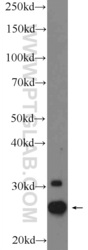
- A431 cells were subjected to SDS PAGE followed by western blot with 25625-1-AP( CHCHD3 Antibody) at dilution of 1:1000
- Sample type
- cell line
- Submitted by
- Proteintech Group (provider)
- Main image
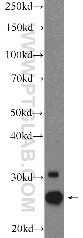
- Experimental details
- The CHCHD3 antibody from Proteintech is a rabbit polyclonal antibody to a fusion protein of human CHCHD3. This antibody recognizes human, mouse antigen. The CHCHD3 antibody has been validated for the following applications: ELISA, WB analysis.

Supportive validation
- Submitted by
- Proteintech Group (provider)
- Main image
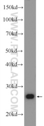
- Experimental details
- mouse liver tissue were subjected to SDS PAGE followed by western blot with 25625-1-AP( CHCHD3 Antibody) at dilution of 1:1000
- Sample type
- tissue
