 Explore
 Validate
 Learn
Learn Western blot
 Immunocytochemistry
Antibody data
- Antibody Data
- Antigen structure
- References [1]
- Comments [0]
- Validations
- Immunocytochemistry [2]
- Flow cytometry [1]
- Other assay [1]
Submit
Validation data
Reference
Comment
Report error
- Product number
- 701058 - Provider product page

- Provider
- Invitrogen Antibodies
- Product name
- Phospho-PRAS40 (Thr246) Recombinant Rabbit Monoclonal Antibody (8H13L13)
- Antibody type
- Monoclonal
- Antigen
- Synthetic peptide
- Description
- Intact IgG appears on a non-reducing gel as ~150 kDa band and upon reduction generating a ~25 kDa light chain band and a ~50 kDa heavy chain. Recombinant rabbit monoclonal antibodies are produced using in vitro expression systems. The expression systems are developed by cloning in the specific antibody DNA sequences from immunoreactive rabbits. Then, individual clones are screened to select the best candidates for production. The advantages of using recombinant rabbit monoclonal antibodies include: better specificity and sensitivity, lot-to-lot consistency, animal origin-free formulations, and broader immunoreactivity to diverse targets due to larger rabbit immune repertoire.
- Reactivity
- Human, Mouse
- Host
- Rabbit
- Isotype
- IgG
- Antibody clone number
- 8H13L13
- Vial size
- 100 μg
- Concentration
- 0.5 mg/mL
- Storage
- Store at 4°C short term. For long term storage, store at -20°C, avoiding freeze/thaw cycles.
Submitted references Secondary Metabolites from the Culture of the Marine-derived Fungus Paradendryphiella salina PC 362H and Evaluation of the Anticancer Activity of Its Metabolite Hyalodendrin.
Dezaire A, Marchand CH, Vallet M, Ferrand N, Chaouch S, Mouray E, Larsen AK, Sabbah M, Lemaire SD, Prado S, Escargueil AE
Marine drugs 2020 Apr 3;18(4)
Marine drugs 2020 Apr 3;18(4)
No comments: Submit comment
Supportive validation
- Submitted by
- Invitrogen Antibodies (provider)
- Main image

- Experimental details
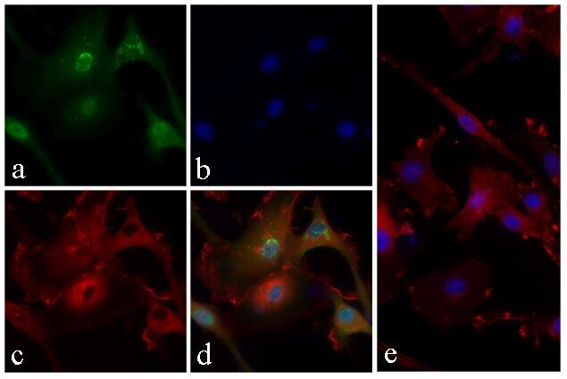
- Immunofluorescent analysis of PRAS40 (pT246) was done by treating serum starved U-87 MG cells with 20 ng/mL PDGF for 15 minutes. The cells were fixed with 4% paraformaldehyde for 15 minutes, permeabilized with 0.25% Triton X-100 for 10 minutes, and blocked with 5% BSA for 1 hour at room temperature. The cells were labeled with PRAS40 (pT246) Recombinant Rabbit Monoclonal Antibody (Product # 701058) at a dilution of 1:400 in 1% BSA and incubated for 3 hours at room temperature and then labeled with Alexa Fluor® 488 Goat anti-Rabbit IgG Secondary Antibody (Product # A-11008) at a dilution of 1:400 for 30 minutes at room temperature (Panel a: green). Nuclei (Panel b: blue) were stained with SlowFade® Gold Antifade Mountant with DAPI (Product # S36938). F-actin (Panel c: red) was stained with Alexa Fluor® 594 Phalloidin (Product # A12381). Panel d is a merged image showing cytoplasmic and nuclear localization. Panel e shows competition with the phospho- PRAS40 (pT246) peptide. The images were captured using a Nikon microscope at 20X magnification.

- Submitted by
- Invitrogen Antibodies (provider)
- Main image

- Experimental details
- Immunofluorescent analysis of PRAS40 (pT246) was done by treating serum starved U-87 MG cells with 20 ng/mL PDGF for 15 minutes. The cells were fixed with 4% paraformaldehyde for 15 minutes, permeabilized with 0.25% Triton X-100 for 10 minutes, and blocked with 5% BSA for 1 hour at room temperature. The cells were labeled with PRAS40 (pT246) Recombinant Rabbit Monoclonal Antibody (Product # 701058) at a dilution of 1:400 in 1% BSA and incubated for 3 hours at room temperature and then labeled with Alexa Fluor® 488 Goat anti-Rabbit IgG Secondary Antibody (Product # A-11008) at a dilution of 1:400 for 30 minutes at room temperature (Panel a: green). Nuclei (Panel b: blue) were stained with SlowFade® Gold Antifade Mountant with DAPI (Product # S36938). F-actin (Panel c: red) was stained with Alexa Fluor® 594 Phalloidin (Product # A12381). Panel d is a merged image showing cytoplasmic and nuclear localization. Panel e shows competition with the phospho- PRAS40 (pT246) peptide. The images were captured using a Nikon microscope at 20X magnification.
Supportive validation
- Submitted by
- Invitrogen Antibodies (provider)
- Main image

- Experimental details
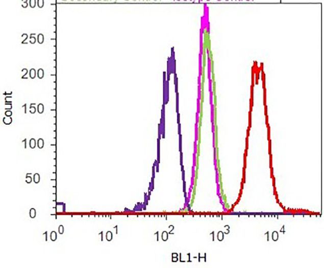
- Flow cytometry analysis of PRAS40 (pT246) was performed on U-87 MG cells treated with PDGF (100 ng/ml, 15 minutes). Cells were fixed with 70% ethanol for 10 minutes, permeabilized with 0. 25% Tritonª X-100 for 20 minutes, and blocked with 5% BSA for 1 hour at room temperature. Cells were labeled with ABfinityª PRAS40 (pT246) recombinant rabbit monoclonal antibody (Product # 701058, red histogram) or with rabbit isotype control (pink histogram) at a dilution of 1:250 in 2.5% BSA. After incubation at room temperature for 3 hours, the cells were labeled with Alexa Fluor¨ 488 goat anti-Rabbit Secondary antibody (Product # A11008) at a dilution of 1:400 for 30 minutes at room temperature. The representative 10,000 cells were acquired and analyzed for each sample using an Attune¨ Acoustic Focusing Cytometer. The purple histogram represents unstained control cells and the green histogram represents no-primary-antibody control.
Supportive validation
- Submitted by
- Invitrogen Antibodies (provider)
- Main image
- Experimental details
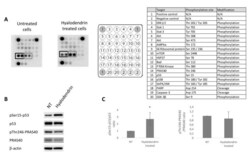
- Figure 4 Intracellular signaling pathways modulated in MCF7-Sh-WISP2 cells exposed to 3 . ( A ) MCF7-Sh-WISP2 cells were treated or not with 2.8 uM of 3 for 48 h. The cells were lysed and the protein extracts were subjected to a protein array analysis. The table chart on the right of the blot indicates the individual proteins tested here. Results are representative of two individual experiments. ( B ) MCF7-Sh-WISP2 cells were treated or not with 2.4 uM of 3 for 72 h. The cells were then lysed and the antigens revealed by immunolabelling using antibodies directed against p53, phospho-p53 (Serine 15), PRAS40 and phospho-PRAS40 (Threonine 246). beta-Actin immunoblot served as a loading control. ( C ) Western blots from three independent experiments were quantified by densitometry and values expressed as (phospho-p53/total p53) or (phospho-PRAS40/total PRAS40) ratios. The statistical analysis of experimental data was performed using Student's paired t-test comparing the hyalodendrin-treated samples with the vehicle control. Results are expressed as means +- standard deviation (SD), * p < 0.05.
